
3-25. DIGITAL IMAGING
Radiology uses a technique called digital imaging to produce higher quality
x-rays. Digital imaging involves the transfer of images composed of dots from one
computer screen to another by digital means. Digital imaging got its start in the 1970s,
with the space program. Incredible images from satellites were produced by
manipulating digital images before photographing them for presentation to the public. In
a similar manner, it is possible to manipulate medical images by computer. This
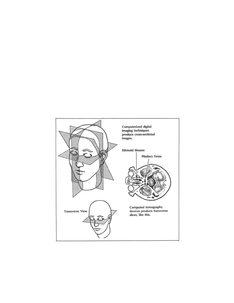
technology reconstructs various physiologic densities to produce layer-by-layer images
of body parts. (See figure 3-11.) The use of dots makes it possible to obtain multiplane
exposures, so that, for example, a radiologist can look at the ethmoid sinuses as well as
the pituitary fossa, immediately posterior. Digital imaging is of great value in diagnosing
patient abnormalities, diseases, and disorders. Digital imaging could make the film
screen cassette, as we know it, obsolete, as resolution and storage problems are
overcome. There are some clinics converting to this system of image storage at the
current time.
Figure 3-11. Computerized imaging produces visual slices of the human body in any
directi

 Previous Page
