
Figure 4-21. Maxillary right lateral incisor.
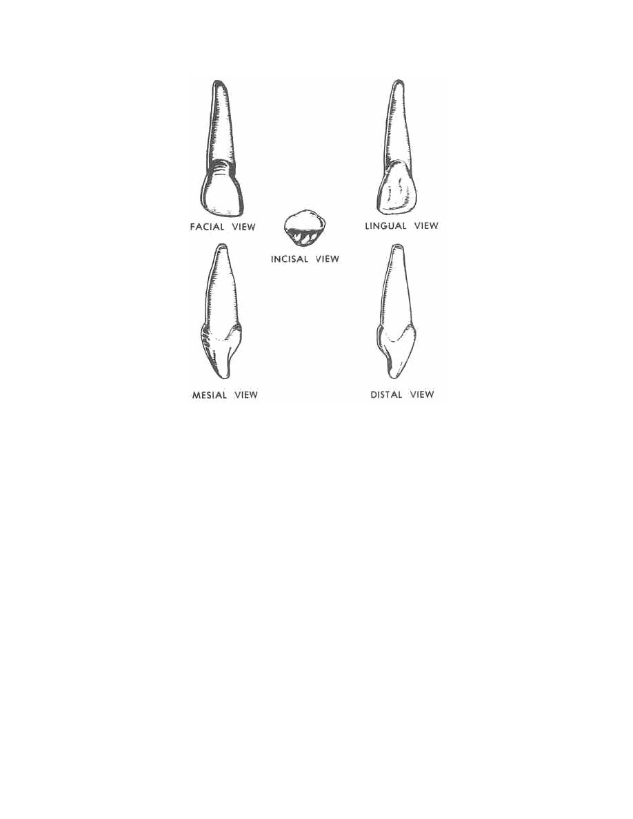
d. Distal Surface. The distal surface is convex in all directions. The distal
contact area is relatively nearer the cervical or gingival margin than in the central
incisor.
e. Incisal Edge. The incisal edge is similar to that of the central incisor. The
outline of the incisal edge reflects generally the greater convexity of the lateral incisor.
f. Root. The root averages about 1 1/2 times the length of the crown. This
single root is smaller than that of the central incisor, but has a greater relative length in
comparison to the length of the crown. It is oval-shaped in cross section. Its apical
one-third inclines toward the distal.
4-12. MAXILLARY CUSPID
The maxillary cuspid (figure 4-22) is the third tooth from the median line. It is the
longest and the only single-cusp tooth in the arch. Located at the angle between the
anterior and the posterior portions of the dental arch, it plays an important role in
determining facial features of the individual and in controlling mandibular movement. It
is sometimes called the "canine tooth" or "eye tooth."
MD0501
4-20

 Previous Page
