
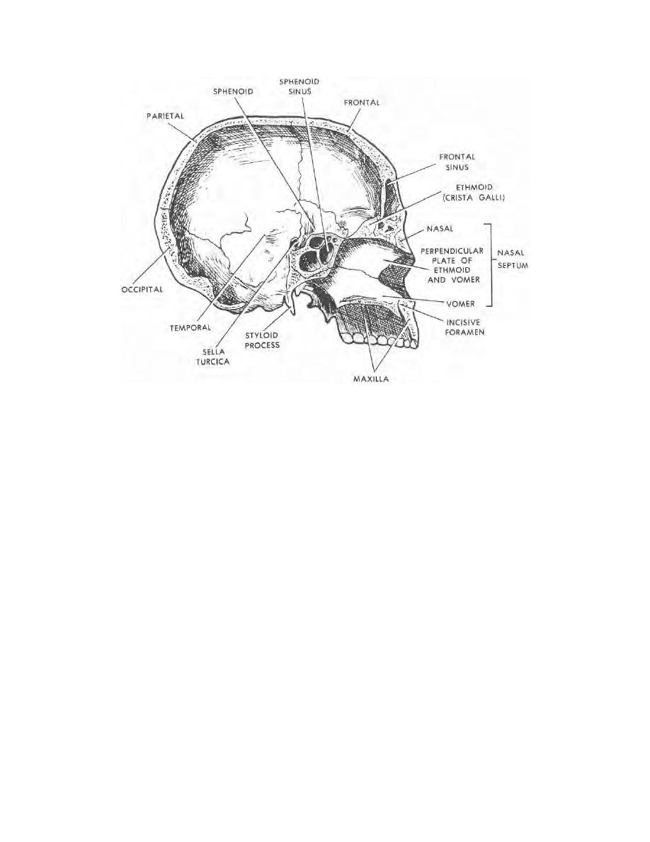
Figure 2-3. Skull (median section).
c. Sphenoid Bone. This bone forms part of the orbits and supports the
posterior part of the upper jaw. It is the central part of the cranium base. The sphenoid
air sinuses lie in the sphenoid bone. The pituitary gland lies in a bony socket called the
sella turcica. The sella turcica is located on the superior aspect of the sphenoid bone.
It transmits the optic nerve (nerve of sight).
d. Ethmoid Bone. This bone makes up the anterior (front) part of the base of
the skull (between the orbits), the medial (toward center) wall of each orbit, part of the
nasal septum, and the roof of the nose. It lies between the eyes and extends from the
frontal bone to the sphenoid bone. The ethmoid bone transmits the olfactory nerve
(nerve of smell).
e. Parietal Bones. These bones form a large part of the cranial vault. They
extend from the frontal bone to the occipital bone. The two bones join at the midline on
top of the cranium. The joining of these bones forms the sagittal suture. From this
suture, these bones extend down and out to near the top of the external ear where they
meet the two temporal bones.
f. Temporal Bones. These two bones form the sides and part of the cranium
base. They contain the organs of hearing and equilibrium. The external acoustic
meatus (an opening or a canal) in the side of each bone forms a passage from the
external ear to the middle ear which lies within each bone.
MD0501
2-4

 Previous Page
