
1-9.
PLEURA
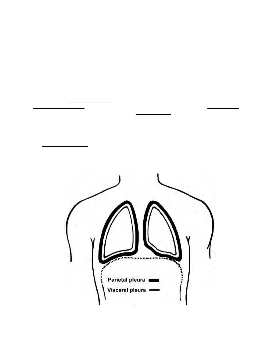
Surrounding each lung individually is a serous cavity called the pleural cavity
(figure 1-9). The minute quantity of serous fluid in the cavity serves as a lubricant. This
serves to minimize friction for the expansion and contraction of the lungs during
breathing.
a. Each lung is covered with a serous membrane called the visceral pleura. The
outer wall of the pleural cavity is lined with another serous membrane known as the
parietal pleura. Areas of the parietal pleura are variously named according to their
location. The mediastinal pleura form the lateral wall of the mediastinum. The
diaphragmatic pleura cover the superior surface of the diaphragm. The costal pleura
line the inner surface of the rib cage. The cupolar pleura form a dome-like extension
into the root of the neck. It contains the apex of the lung.
b. When each lung is in its smaller volume, its corresponding diaphragmatic
pleura lies close to the lower costal pleura. The slit-like cavity between them is called
the costophrenic sinus. Fluids of each pleural cavity tend to collect in this sinus since it
is the lowest area for each. When the diaphragm contracts and flattens out, each
costophrenic sinus opens up, and the inferior portion of the expanding lung occupies
this space.
Figure 1-9. Lungs, visceral, and parietal pleura.
MD0568
1-12

 Previous Page
