
b. Removal of Foreign Body. This is done very gently using aseptic technique
to avoid secondary infection.
(1) Serious damage to the ocular structures often results from the careless
or unskilled removal of foreign bodies from the eye.
(2) The foreign bodies which most commonly cause injury and irritation of
the conjunctiva or cornea are dust particles from grinding wheels, cinders, street dirt,
gravel, and grains of sand. Foreign bodies such as splinters of wood, metal, or glass
which become embedded in or penetrate the eye often cause serious damage.
(3) A foreign body, which is lying on the cornea, is embedded in, or
penetrates the eye, is always removed by a medical officer.
c. Graft of Cornea. Opaque corneal tissue is excised and healthy corneal
tissue of the same size and shape is placed. The operation is done to restore vision by
permitting light to enter the eye. An important factor in the success of this surgery is
that the donor tissue absolutely be fresh. If opacity (the condition in which light cannot
penetrate) has begun to develop in the graft tissue, the success of the operation is
doubtful. Eye "banks," similar to other tissue "banks," provide for acquisition,
preservation, and transportation of healthy corneal tissue to the hospital where the
operation is to be done.
Section II. EAR SURGERY
1-18. GENERAL ANATOMY AND PHYSIOLOGY OF THE EAR
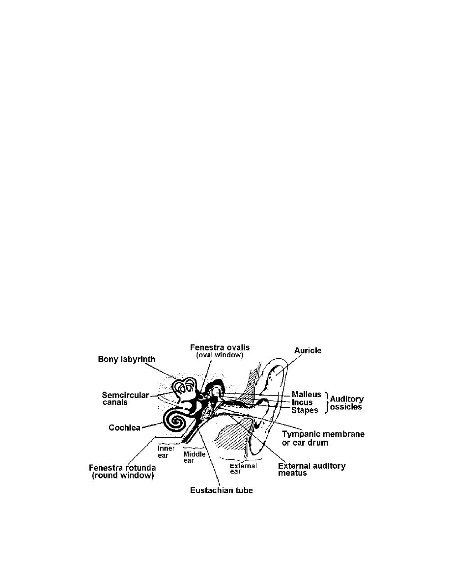
The ear (see figure 1-5) is made up of three distinct divisions: the external ear,
the middle ear, and the inner ear. The middle and inner ear structures are situated in
the temporal bone cavity.
Figure 1-5. Parts of the ear
MD0928
1-19

 Previous Page
