
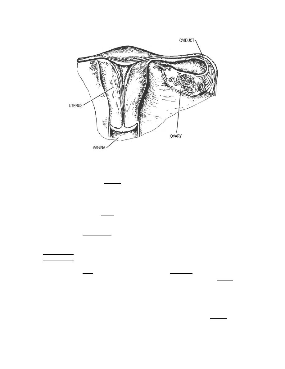
Figure 3-13. Frontal section of the female reproductive organs showing an oviduct and
the pear-shaped uterus.
d. The Vagina. The vagina (figures 3-12 and 3-13) is a thin-walled, tubular
organ extending from the cervix of the uterus to the vulva where it opens to the exterior.
It averages 8 centimeters in length and lies dorsal to the bladder and urethra-ventral to
the rectum. The vagina is also part of the birth canal.
e. The Vulva. The vulva (external genitalia) includes the mons pubis, labia
majora, labia minora, clitoris, vestibule, and the greater vestibular glands (figure 3-14).
(1) The mons pubis is the rounded eminence anterior to the symphysis
pubis, consisting mainly of fibrous and adipose tissue. Extending downward from the
mons pubis and backwards toward the anus are two longitudinal cutaneous folds called
the labia majora. Within these two folds of skin are two smaller cutaneous folds, called
the labia minora, which meet anteriorly and form the prepuce of the clitoris.
(2) The cleft between the labia minor is the vestibule, which contains the
clitoris, the urethral and vaginal orifices, and many vestibular glands. The clitoris, the
homologue of the male penis, is a small body of erectile tissue located at the point
where the two labia minor meet. The urethral orifice is located about 2.5 centimeters
posterior to the clitoris and immediately anterior to the vaginal orifice. The urethra has
no connection with the reproductive system. The urethra conveys urine. The vaginal
orifice occupies about two-thirds of the posterior region of the vestibule and is separated
from the cavity of the vestibule by a thin fold of mucous membrane, the hymen. The
hymen may completely cover the vaginal orifice (imperforate hymen) or it may be
absent entirely.
MD0956
3-24

 Previous Page
