
LESSON 4
SPECIAL RADIOGRAPHIC PROCEDURES
Section I. MAMMOGRAPHY
4-1.
INTRODUCTION
Radiographic examination of the female and male breast is known as
mammography. This examination is used for the investigation of pathological
symptoms to detect the nature and causes for those symptoms, to differentiate between
benign and malignant tumors, and to determine a course of treatment.
4-2.
ANATOMY AND PHYSIOLOGY OF THE BREAST
Functionally, the female breasts are accessory glands of the reproductive
system. Several types of tissue occur in varying amounts, depending upon the age and
physical condition of the patient.
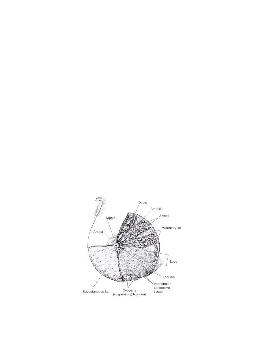
a. The External Structure. The surface components of the breast include the
skin, the areola, and the nipple. Usually, the breasts extend from about the second rib
to the sixth or even seventh rib and from the lateral margins of the sternum to the axilla.
Although breasts differ in size, the average craniocaudad (vertical) diameter is between
12 to 14 cm while the transverse (horizontal) diameter is slightly larger. In figure 4-1,
note the structures of the breast, specifically the areola and the nipple. The nipple is a
projection of areolar tissue on the apex of the breast. Its tip is perforated by 15 to 20
tiny orifices to the lactiferous tubules.
Figure 4-1. Breast diagram showing the breast structures.
MD0959
4-2

 Previous Page
