
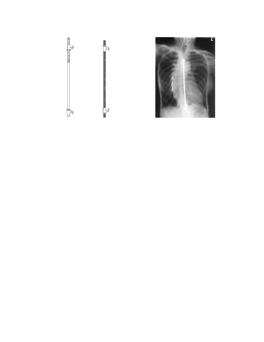
Figure 4-11: Harrington rods: Diagram on left, radiographic on right.
Section VI. RADIOGRAPHIC DEMONSTRATION OF FLUID LEVELS
4-37. INTRODUCTION
The collection, dispersion, shifting, or superimposition of free fluid with
contiguous or ambient structures within the body cavities often requires a special
technique for adequate diagnostic demonstration, such as differentiation between free
fluid and thickened membranes or determination of the amount and behavior of free
fluid within a body cavity. The procedure by which this is accomplished is known as
fluid-level radiograph. The regions most commonly examined are the paranasal
sinuses, the interpleural spaces, and the abdominal cavity.
4-38. PRINCIPLES
a. There is one prime requisite that must remain constant at all times when
performing fluid-level radiography--the CR (or projection) must always be horizontal.
Also, as nearly as circumstances permit, the horizontal CR should be parallel with, and
at the same elevation as, the plane of the fluid level.
(1) Figure 4-12, part A, shows the horizontal CR at the same elevation as
the plane of the fluid level. This demonstrates the plane of the fluid level with clear
demarcation.
(2) Figure 4-12, part B, shows the effect of aligning the horizontal CR at a
lower elevation in relation to the fluid level, the actual projection being accomplished by
vertical divergent rays origination from the same source-point as the horizontal CR. The
resultant image demonstrates a distorted and diffused outline of the fluid level which, in
some cases, may be of doubtful diagnostic value.
MD0959
4-29

 Previous Page
