
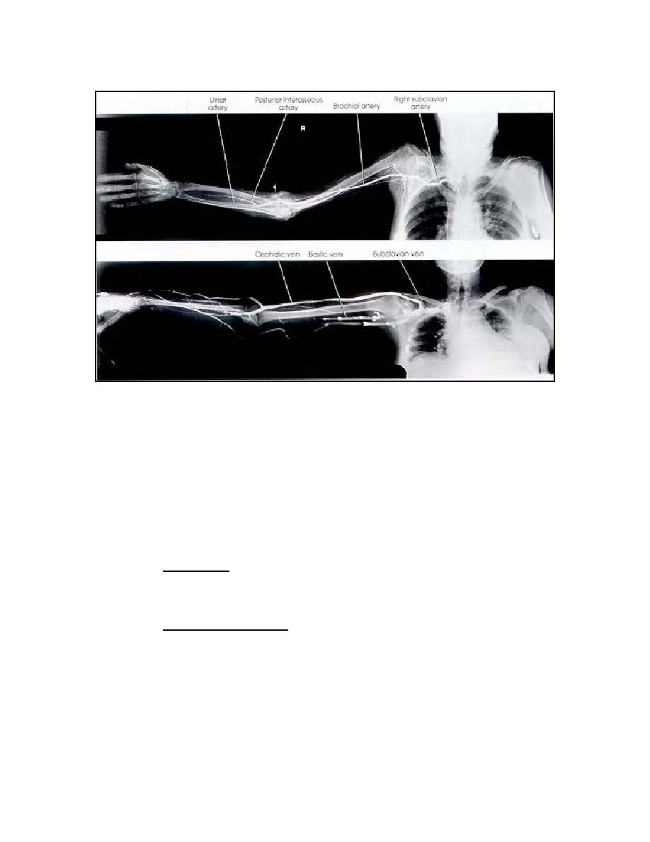
The top projection demonstrates the right subclavian artery injected,
demonstrating iatrogenic occlusion of radial artery (arrow).
The bottom is a normal right upper venogram.
Figure 3-8. Venography.
3-20. ANGIOGRAPHIC EXAMINATION OF THE LOWER EXTREMITIES
a. Arteriography.
(1) Preparation. Digital subtraction is used to demonstrate any part of the
lower extremity. Patient preparation and administration of sensitivity test is covered
earlier.
(2)
Preliminary procedure.
(a) As soon as the patient is received at the x-ray clinic, the specialist
will ascertain from the examiner the area(s) of the extremity to be examined. If the
examination is concerned with any part lying distal to the region of the knee, then non-
grid technique is normally employed. Bucky technique may be indicated when the
arterial channels in the thigh region (exclusively) or in the leg and thigh regions are to
be examined. With the latter, it is possible to obtain simultaneous coverage of both the
leg and thigh portions with a single exposure, by using several cassettes (placed end-
MD0959
3-21

 Previous Page
