
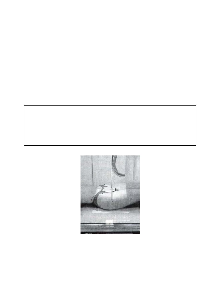
e. The Conefield. The fourth and final table factor is the conefield (CF). The
conefield or area of the beam of radiation striking the film must be reduced to only that
area required to produce the view requested. By adjusting the conefield setting, the
size of the radiation field is set (figure 2-17). You may specify a conefield of 4 inches, 6
inches, 8 inches, or any other size, as required, to produce the desired view.
Collimators made of lead or other metal with high radiation absorption power are used
to restrict the conefield by so confining the beam. The peripheral (extra) radiation
strikes and is absorbed by the intervening metal while only the rays in line with the exit
aperture are transmitted to the exposure field. Or, you may opt for a full field coverage
(FFC), that is, no coning down to limit the area of exposure. This may be appropriate
for specific visualizations of the anatomy. Most views will require full field coverage
(FFC), that is, if half the cassette is used, then the beam of radiation covers the entire
half. If the whole cassette is used, the beam covers the entire cassette and there is no
restricted conefield. Other views, such as those of the sinuses and the cervical spine
will require a restricted conefield.
conefield: the area of the beam of radiation striking the film.
full field coverage (FFC): total exposure of the area of the film cassette that is used:
no coning down.
Figure 2-17. The patient is positioned with the conefield set.
MD0961
2-23

 Previous Page
