
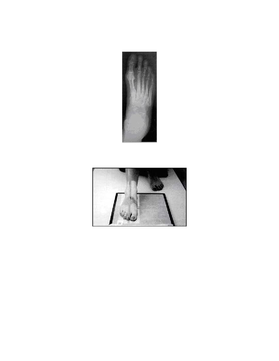
b. Radiographic Image. The radiographic image displays the correct
anatomical structures of a properly positioned body part.
c. The Patient Positioning Photograph. This image depicts proper positioning
of the patient, in this case positioning for an AP foot.
d. The Order of Procedure Chart. The information generally included in the
order of procedure chart is explained in the next two figures. Note that items a, b, and c
are the same for all positions, that is, no position specific or unique information is
provided for these entries. They will depend on the anatomy that needs to be
demonstrated. Figure 1-1 explains the order of procedure chart.
MD0962
1-3

 Previous Page
