
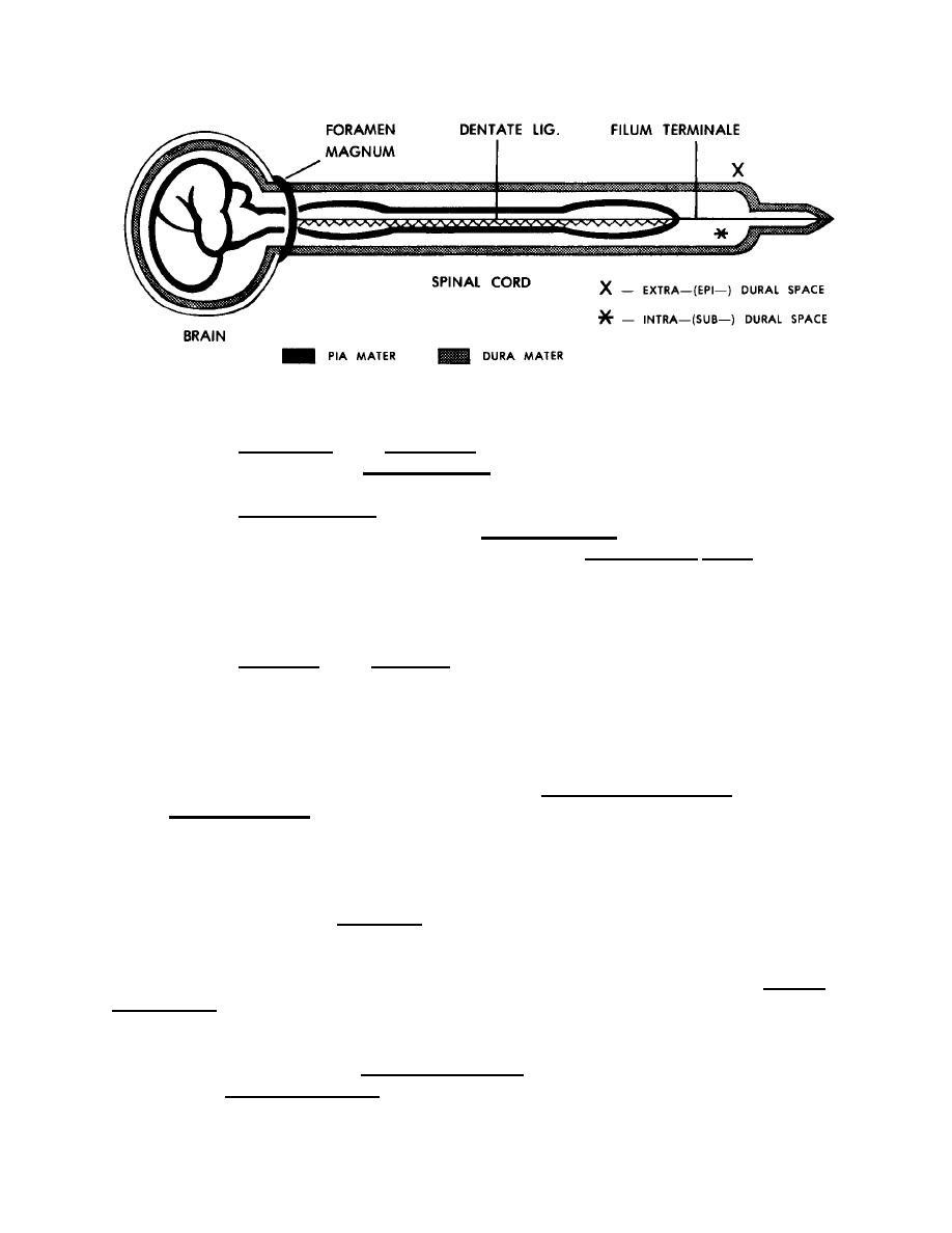
Figure 11-7. A schematic diagram of the meninges, as seen in side view of the CNS.
(1) Dura mater. The dura mater is a tough outer covering for the CNS.
Beneath the dura mater is the subdural space, which contains a thin film of fluid.
(2) Arachnoid mater. To the inner side of the dura mater and subdural
space is a fine membranous layer called the arachnoid mater. It has fine
spiderweb-type threads which extend inward through the subarachnoid space to the pia
mater. The subarachnoid space is filled with cerebrospinal fluid (CSF).
ARACHNOID = spider-like
(3) Pia mater. The pia mater is a delicate membrane applied directly to the
surface of the brain and the spinal cord. It carries a network of blood vessels to supply
the nervous tissues of the CNS.
11-12. BLOOD SUPPLY OF THE CNS
a. Blood Supply of the Brain. The paired internal carotid arteries and the
paired vertebral arteries supply blood rich in oxygen to the brain. Branches of these
arteries join to form a circle under the base of the brain. This is called the cerebral circle
(of Willis). From this circle, numerous branches supply specific areas of the brain.
(1) A single branch is often the only blood supply to that particular area.
Such an artery is called an end artery. If it fails to supply blood to that specific area, that
area will die (stroke).
(2) The veins and venous sinuses of the brain drain into the paired internal
jugular veins, which carry the blood back toward the heart.
b. Blood Supply of the Spinal Cord. The blood supply of the spinal cord is by
way of a combination of three longitudinal arteries running along its length and
reinforced by segmental arteries from the sides.
MD0006
11-14

 Previous Page
