
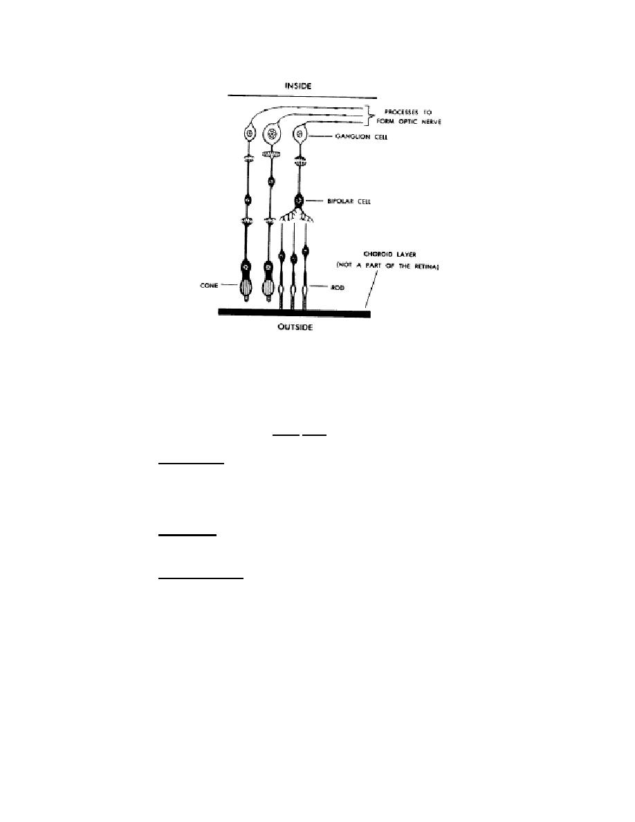
Figure 5-2. Cellular detail of retina.
(c) Associated with the rods and cones are the beginnings of neurons
of the optic nerve. These neurons pass out of the bulbus oculi at the posterior end (in a
point medial and superior to the fovea centralis). At the point of exit, there are not rods
or cones. Therefore, it is called the blind spot (optic papilla/optic disk).
(2) Ciliary body. The anterior end of the choroid layer thickens to form a
circular "picture frame" around the lens of the bulbus oculi. This is also near the margin
of the base of the cornea. The frame-like structure is called the ciliary body. It includes
mostly radial muscle fibers, which form the ciliary muscle.
(3) Ligaments. The lens is suspended in place by ligaments. These
ligaments connect the margin (equator) of the lens with the ciliary body.
(4) Crystalline lens. The crystalline lens is located in the center of the
anterior of the bulbus oculi, just behind the cornea.
(a) The lens is biconvex. This means that it has two outwardly curved
surfaces. The anterior surface is flatter (less curved) than the posterior surface.
(b) The lens is transparent and elastic. As one grows older, the lens
becomes less and less elastic. The ligaments maintain a tension upon the lens. This
tension keeps the lens flatter and allows the lens to focus on distant objects. When the
ciliary muscle contracts, the tension on the lens is decreased. The decreased tension
allows the lens to thicken. The greater thickness increases the anterior curvature and
allows close objects to be seen clearly.
MD0805
5-4

 Previous Page
