
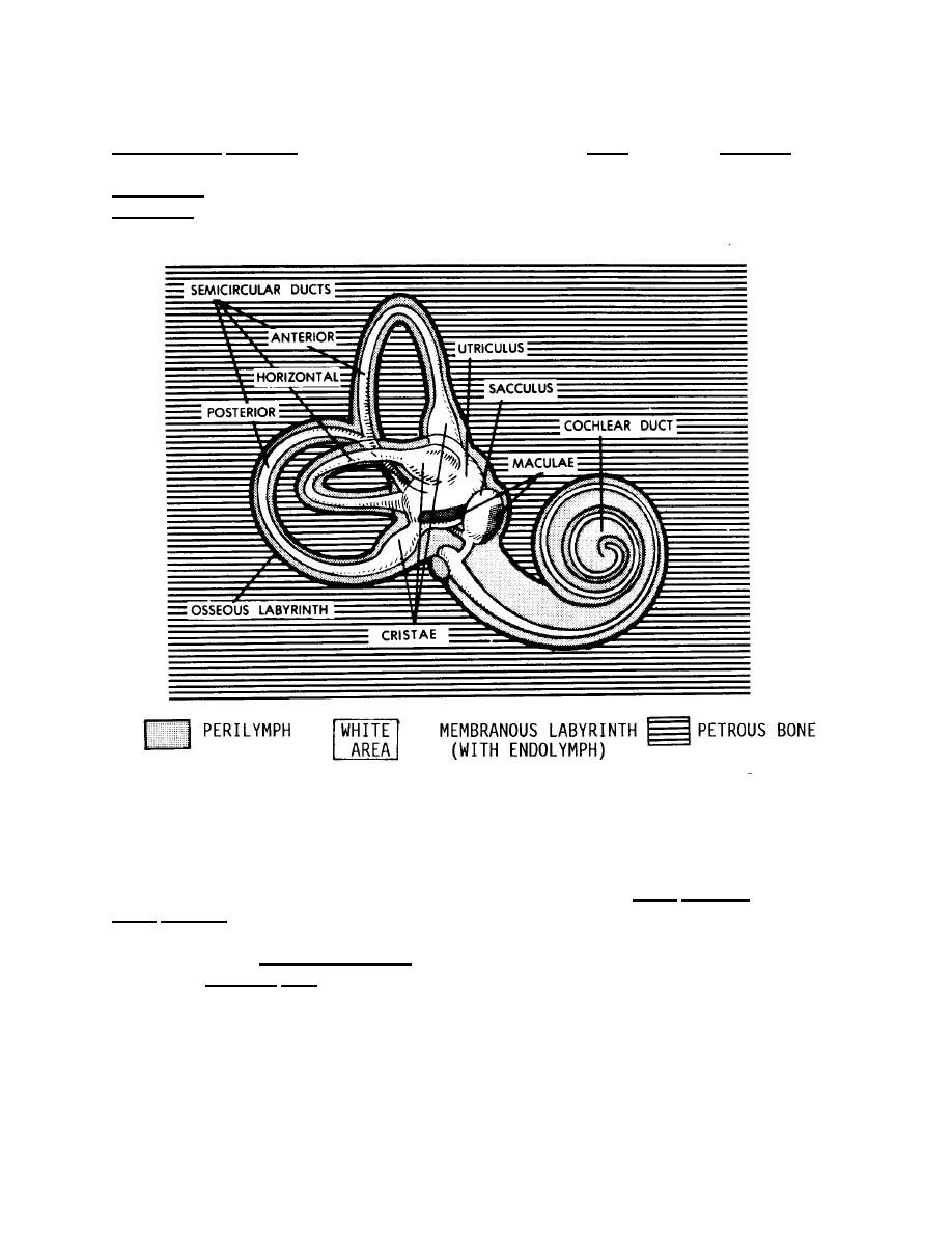
b. Organization of the Internal Ear. The internal ear is essentially a
membranous labyrinth suspended within the cavity of the bony (osseous) labyrinth of
the petrous bone (Figure 13-8). The membranous labyrinth is filled with a fluid, the
endolymph. Between the membranous labyrinth and the bony labyrinth is the
perilymph.
Figure 13-8. The labyrinths of the internal ear.
c. The Cochlea. The cochlea is a spiral structure associated with hearing.
Its outer boundaries are formed by the snail-shaped portion of the bony labyrinth. The
extensions of the bony labyrinth into the cochlea are called the scala vestibuli and the
scala tympani (Figure 13-7B). These extensions are filled with perilymph.
(1) Basilar membrane (Figure 13-7B). The basilar membrane forms the
floor of the cochlear duct, the spiral portion of the membranous labyrinth. The basilar
membrane is made up of transverse fibers. Each fiber is of a different length, and the
lengths increase from one end to the other. Thus, the basilar membrane is constructed
similarly to a harp or piano. Acting like the strings of the instrument, the individual fibers
mechanically vibrate in response to specific frequencies of pulses in the perilymph.
Thus, each vibration frequency of the sound stimulus affects a specific location of the
basilar membrane.
MD0007
13-14

 Previous Page
