
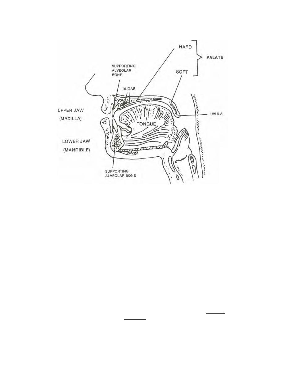
Figure 3-1. Cross-section of the mouth.
c. Opening of the Parotid Gland. The parotid gland, the largest of the salivary
glands empties its contents (saliva) into the oral vestibule through an opening called
Stensen's duct (which is another name for the parotid duct). See figure 2-17. The
opening can be found opposite the crown of the maxillary second molar and is generally
marked by a high elevation of mucous membrane, which is the parotid papilla.
d. The Teeth. When the mouth is open wide and the jaws are apart, the teeth
can be seen to be arranged in arches with open ends directed backwards or posteriorly.
The maxillary teeth are in the maxilla or upper jaw. See figure 3-2. The mandibular
teeth are in the mandible or lower jaw. See figure 3-3. If an imaginary vertical line (the
midline) is drawn between the central incisors and extended backward, it will cut each
arch into two halves, one the mirror image of the other. Each of these parts is termed a
quadrant. Thus, there is a maxillary right and a maxillary left quadrant and a mandibular
right and a mandibular left quadrant. There are eight permanent teeth in each quadrant.
Viewing each quadrant from the midline posteriorly, the two incisors (the central and
lateral), one cuspid, two bicuspids (premolars), and three molars make up the
arrangement of the teeth. The incisors and cuspids are known as the anterior teeth,
and the bicuspids and molars as the posterior teeth.
MD0501
3-3

 Previous Page
