
(2) Scales. Scales are a buildup of dry cells (horny layer) that is higher than
usual. Although there are a large variety of scales, some are so distinctive that they can
be used to diagnose specific skin problems. For example, a chronic plaque-like scale
that is silvery-white to gray is usually psoriasis. Greasy, yellowish scales may indicate
seborrheic dermatitis. If the scales are dry and diffuse and look like fish scales on the
lower legs, the skin disease is icthyosis. Skin lesions of pityriasis rosea and tinea corpis
scale mainly at the edge of individual lesions.
(3) Fissure. A fissure is a crack in the epidermis extending into the dermis.
They are linear cleavages in the skin, sometimes very painful. These cracks occur
particularly in the hands and feet, especially after therapy has caused excessive drying
of the skin. Fissures also occur at the angles of the mouth.
(4) Erosion. A loss of epidermis that does not extend into the dermis is
termed erosion. Erosion is often seen in herpes infections.
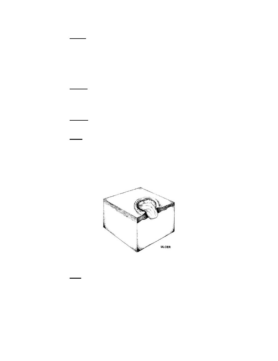
(5) Ulcer. An ulcer (figure 3-11) is a skin lesion in which there has been
destruction of the epidermis and the upper papillary dermis. An ulcer always results in a
scar. To determine the cause of an ulcer, note the ulcer's location, borders, base,
discharge, and any associated features of the lesion such as nodules, excoriations
(scratch marks), varicosities (abnormal swellings), hair distribution, presence or
absence of sweating, and adjacent pulses. There are many causes of ulcers--skin
trauma of all kinds (heat, cold, electrical, chemical); bacterial, viral, and fungal
infections; parasitic infestations; and tumors to name just a few.
Figure 3-11. Ulcer.
(6) Scar. A scar (figure 3-12) is a fibrous healing of a wound, healing that
replaces the normal dermis and epidermis that have been damaged. Scars in different
areas of the body may look different. Hypertrophic scars (scars with excessive fibers)
result when a lot of collagen is produced in the healing process. This type of scar may
occur in the course of acne, herpes zoster, and porphyria (a disorder of blood pigment
metabolism). In atrophic scars, the epidermis is thin and usually has neither skin lines
nor appendages. This type of scar may be depressed.
MD0575
3-9

 Previous Page
