
Section IV. MICROSCOPY
1-23. INTRODUCTION
A modern binocular microscope is essential for the study of parasites. A
compound microscope uses a combination of lenses (i.e., the objective and the ocular)
to magnify the object. The objective lenses projects an enlarged primary image near
the top of the tubular barrel. This aerial image is further magnified by the ocular lens,
which projects it to the retina. The final image at the retina is called the virtual image.
Through the compound microscope, this image is projected in a phase contrast system
that allows for the differences that occur between light altered by the object and the
unaltered (or background) light. The application of this principle (as provided by Kohler)
gives a higher quality of resolution in observing cellular phenomena.
1-24. EQUIPMENT
A microscope (see figure 1-1) properly equipped with a lens system, an illumination
system, a condenser, filters, a diaphragm, a prism, and a mechanical stage is suitable
for diagnostic parasitology.
a. The Lens System. The lens system consists of the oculars (eye pieces) and
the objectives.
(1) The oculars (eye pieces). In general, microscopes used in parasitology
are provided with ten power (10X) wide field oculars.
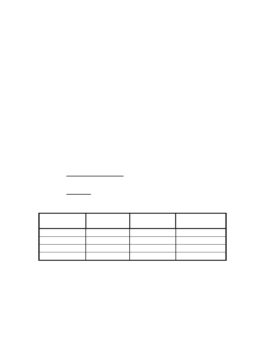
(2) Objectives. Microscopes have a rotating nosepiece to which three or
four objectives are attached. These objectives are of different magnifications, different
working distances, and are distinguished by a color band.
FOCAL
OBJECTIVE
COLOR BAND
MAGNIFICATION
DISTANCE
Scanner
Black
7.2 mm
4X
Low power
Green
4.3 mm
10X
High power
Yellow
0.7 mm
45X
Oil immersion
Red
0.1 mm
100X
b. Magnification. By multiplying the power of the ocular (10X) by the power of
the objective (4X, 10X, 45X, or 100X), total magnification (40X, 100X, 450X, or 1000X)
is obtained.
MD0841
1-18

 Previous Page
