
(3) Cytoplasm. The cytoplasm contains many small, evenly distributed light
pink granules.
g. Neutrophilic Segmented Cell.
Size. Ten to 16 microns in diameter.
(1)
(2) Nucleus. The nucleus has definite lobes separated by a very narrow
filament or strand. The nucleus-cytoplasm ratio is approximately 1: 3.
(3) Cytoplasm. The cytoplasm is light pink and the small, numerous, and
evenly distributed neutrophilic granules have a light pink color.
h. Development of the Eosinophilic Group. Cells of the eosinophilic group
are characterized by relatively large, spherical, cytoplasmic granules that have a
particular affinity for the eosin stain. The earliest eosinophil (myelocyte) has a few dark
spherical granules with reddish tints that develop among the dark, nonspecific granules.
As the eosinophilic cells pass through their various developmental stages, these
granules become less purplish-red and more reddish-orange. The dark blue,
nonspecific granules, characteristic of the promyelocyte and the early myelocyte stages,
disappear. Because the percentage of eosinophils is usually low in bone marrow
peripheral blood smears, no useful clinical purpose is served by routinely separating the
eosinophils into their various myelocyte, metamyelocyte, band, and segmented
categories. On the other hand, in situations such as eosinophilic leukemia in which the
eosinophils are greatly increased, an analysis of the incidence of the various stages
would be useful in diagnosis.
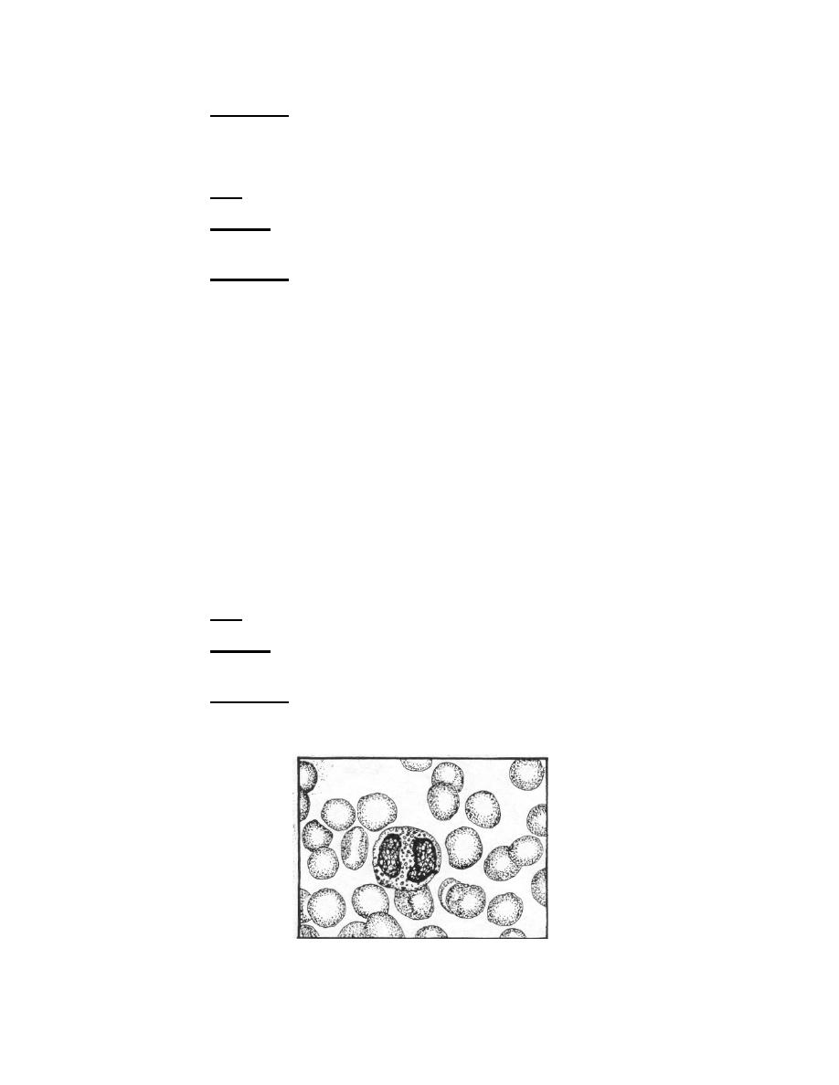
i. Mature Eosinophil.
Size. Ten to 15 microns in diameter.
(I)
(2) Nucleus. The nucleus has definite lobes separated by a very narrow
filament or strand. Seldom does an eosinophil have more than two lobes.
(3) Cytoplasm. The cytoplasm contains bright reddish-orange, distinct
granules. The granules are spherical, uniform in size, and evenly distributed throughout
the cytoplasm, but rarely overlie the nucleus.
Figure 4-3e. Granulocytic series: Eosinophil.
MD0853
4-15

 Previous Page
