
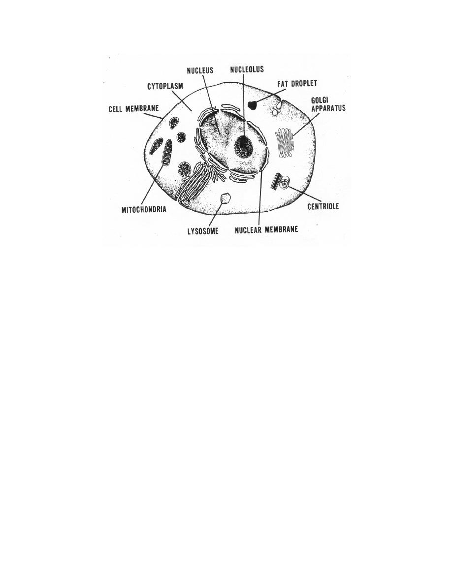
Figure 1-1. Typical cell structure.
within the nucleus is a network of nuclear fibrils made up of DNA (deoxyribonucleic
acid) and protein called chromatin. It is thought that the decreasing growth activity of a
cell during maturation is regulated by chromatin.
c. Surrounding the nucleus is a mass of protoplasm called cytoplasm.
Contained within the cytoplasm are numerous granules, filaments, and globules. These
structures are divided into two groups known as organoids (organelles) and inclusions.
The organoids are thought to perform most of the metabolic functions of the cell.
Mitochondria, Golgi apparatus, fibrils, centrioles, and the chromatin substance are
classified as orgnanelles. Cytoplasm inclusions are usually seen as granulation. The
granulation is an accumulation of proteins, lipids, carbohydrates, pigments, and
secretory granules.
1-3.
CELLULAR CONSTITUENTS
a. Erythrocytes. An erythrocyte (red blood cell) is an elastic, non-nucleated,
biconcave disc having a diameter of approximately 7.2 microns. The mature red cell
contains about 34 percent hemoglobin (a complex iron-bearing pigment that transports
oxygen). Hemoglobin is contained in the interior of the cell, and the outer surface of the
cell is surrounded by a cell membrane. When unstained, the cell has a pale, greenish-
yellowish appearance. It is buff pink with an accented central zone of pallor when
stained with Wright's stain. The production of erythrocytes or erythropoiesis, occur
primarily in the red marrow of the spongy bones. Erythrocytes make up the great
majority of cells found in the peripheral blood. Their vast surface area is important in
the transport of oxygen from the lungs to the tissues because of quick exchange of
oxygen in both sites that occurs across the red cell surface. Erythrocytes are subject to
MD0853
1-3

 Previous Page
