
(3) Cytoplasm. The cytoplasm stains a bluish-buff with Wright's stain and
there is no central light pallor as in the erythrocyte. With supravital staining, this cell will
show light blue reticulum strands in the cytoplasm.
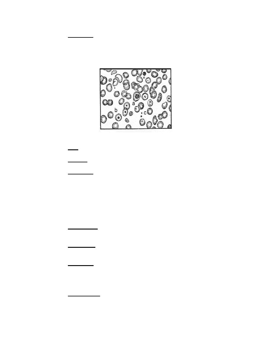
f. Erythrocyte.
Figure 4-1f. Erythrocytes series: Erythrocyte.
Size. Six to 8 microns in diameter.
(1)
(2)
Nucleus. The nucleus is absent.
(3) Cytoplasm. The cytoplasm of the periphery is light orange with a central
zone of pallor. The appearance of the central zone of pallor is due to the biconcave
morphology of the cell, which allows more light through the center than through the
margin areas.
4-5.
VARIATIONS IN ERYTHROCYTES
a. Size.
(1) Anisocytosis. Anisocytosis is a variation in the size of erythrocytes
beyond the normal limits. Cells of varying size are seen in the same fields.
(2) Macrocytes. Macrocytes are erythrocytes larger than nine microns in
diameter. These cells may be found in liver disease.
(3) Microcytes. These erythrocytes are smaller than 6 microns in diameter.
These cells are found in thalassemia and other anemias.
b. Shape.
(1) Poikilocytosis. This term describes a marked variation in the shape of
erythrocytes. Poikilocytes can be pear-shaped, comma-shaped, oval- shaped, or
various other bizarre forms. These cells are encountered in pernicious anemia and
many other types of anemia.
MD0853
4-6

 Previous Page
