
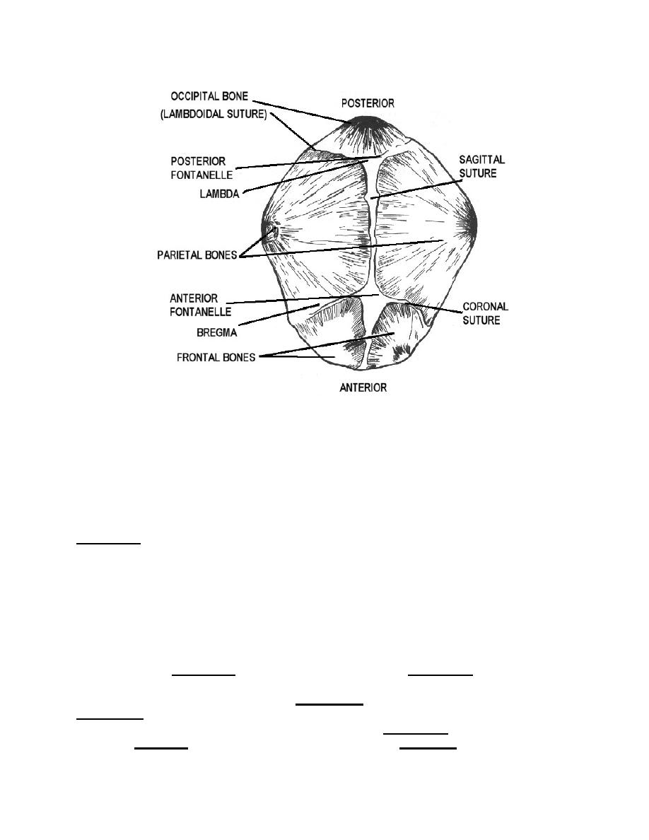
Figure 1-14. Anterior and posterior fontanelles, infant's skull.
b. Cartilaginous (Endochondral) Formation. The tones of the rest of the
skeleton are preformed in cartilage. Ossification proceeds from an ossification center
toward the extremities, which remain cartilaginous for some time. Subsequently, a
similar process begins in one or more places in the extremities and gradually proceeds
toward the center. An area of cartilaginous tissue persists after birth for various periods
of time. In infants and children, this area affords growth in length. It is called the
epiphyseal zone (the suffix "physeal" means "growth").
c. Cranial Growth. Growth of the cranial bones is affected in formative steps,
which are modifications of intramembranous formation. Their development entails
ossification of the membranous fontanelles that becomes complete when the child is
approximately 2 years old.
d. Growth of Other Bones. The remaining bones of the skeleton undergo
changes similar to those of a long bone during growth, that is, an increase in diameter
and length. The periosteum that covers the bone contains osteoblasts that
progressively deposit layers of bone to form the external portion of the bone. Correlated
with this growth in diameter externally, osteoclasts (bone-destroying cells) in the
endosteum destroy some of the bone internally, thereby enlarging the internal
(medullary) canal. Growth in length takes place in the epiphyseal zones. The shaft is
called the diaphysis and the end of the bone is called the epiphysis (figure 1-11).
MD0956
1-24

 Previous Page
