
b. When it is necessary or desirable to demonstrate multiple fluid levels (situated
at different elevations in relation to the horizontal CR) on a single radiograph (for
example, the abdominal region), increasing the SID will tend to obviate some of the
adverse effects of "off-center" projection.
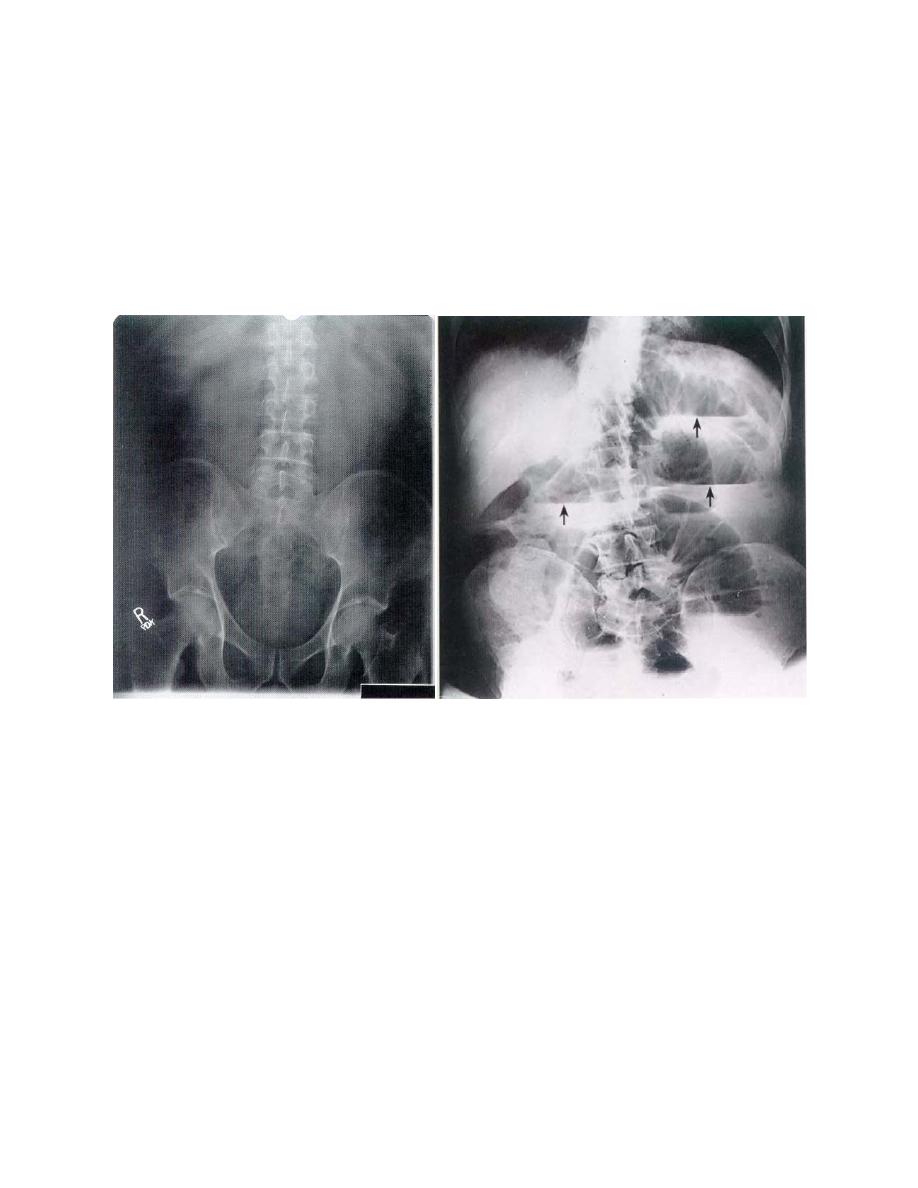
c. In general, the behavior of free fluid is demonstrated radiographically in
figures 4-13 (exposed with CR in vertical position) and 4-14 (exposed with CR in
horizontal position). Notice in figure 4-13, there is no evidence of the disposition of fluid,
while in figure 4-14; the fluid is in well-defined levels.
Figure 4-13. Projection of abdomen
Figure 4-14.
Projection of abdomen made
made with the central ray in the
with the central ray in the horizontal
vertical relationship and the patient in
relationship and the patient in the erect
the supine position. This view shows
position. Note disposition of fluid into well-
no definite evidence of the disposition
defined levels.
of fluid.
4-39
CLINICAL PROCEDURE
a. After the patient has been placed in the position in which the radiograph is to
be made, it is generally advisable to allow an elapse of 2 to 4 minutes before making
the exposure. This interval permits gravitation or "settling" of the free fluid
b. Fluid-level radiography can be performed with the patient in the erect, supine,
prone, or lateral decubitus position or with the patient placed in various inclined-plane
positions, depending upon clinical desires or dictates (figures 4-15 and 4-16).
MD0959
4-31

 Previous Page
