
(2) Preliminary films of the skull (AP and lateral) are exposed according to
the examiner's instructions. These films are processed immediately and presented to
the examiner for reading.
(3) A sterile layout consisting of the instruments and materials required for
cerebral arteriography is prepared.
(4) Radiation protection shielding should be arranged so as to provide the
necessary safety and yet afford optimum freedom of movement for each member of the
medical team.
(5) If the injection is to be carried out by the percutaneous method, the
examiner palpates the femoral vein in the groin area to determine the best site for
puncture.
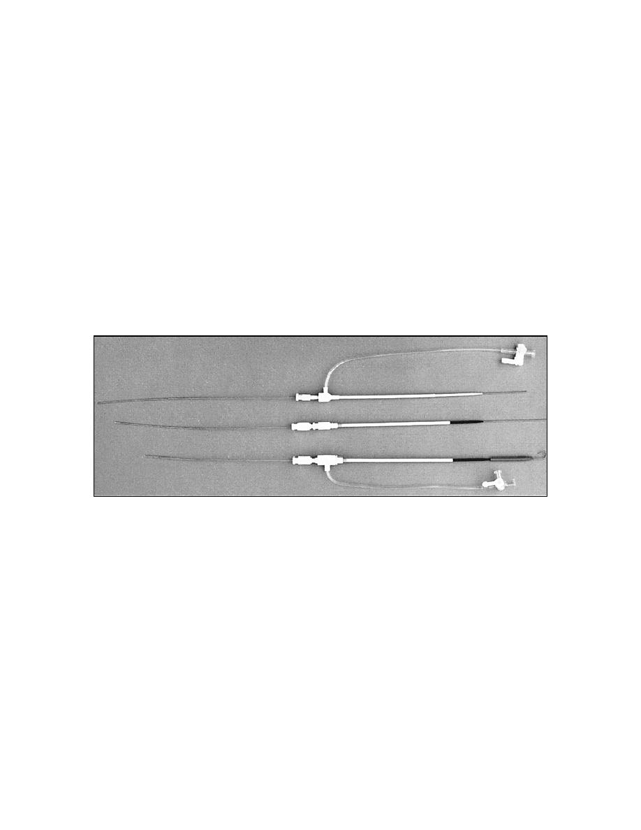
(6) The examiner inserts the injection needle into the selected lumen of the
vein and then engages the needle with the adapter in the injection system (figure 3-14).
Figure 3-14. Showing relationships of the inserted injection needle, adapter,
syringe, and stopcock complex for selectively directing the flow of
contrast medium in the injection continuum.
c. Injection of Contrast Medium and Radiography. See figures 3-15 and
3-16.
(1) The specialist adjusts the patient's head in the AP position. The CR is
aligned parallel to the glabellomeatal line and directed to a point midway on line
approximately 2 cm superior to the tragi.
(2) A syringe filled with approximately 20 cc of warmed contrast solution is
attached to the stopcock complex.
MD0959
3-38

 Previous Page
