
(1) The large breast usually requires both a larger cone field and a decrease
in technique. By increasing the SID over that used for the medium breast, both of the
requirements are met.
(2) Two exceptions to this method are the small breast containing mostly
fatty tissue and the large breast containing mostly a fibroglandular tissue. These are
usually identified by the x-ray specialist before the examination and appropriate
compensations are made.
4-9.
PATIENT POSITIONING
The projections most commonly used for mammography studies are:
craniocaudad, mediolateral, and axillary.
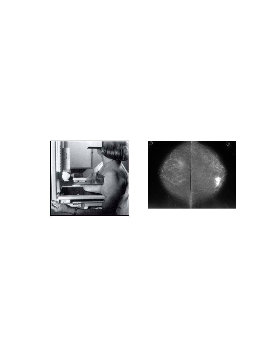
a. Craniocaudad. In figure 4-3, the major considerations associated with the
craniocaudad projection are illustrated. When positioning a patient for this projection,
use the following as a guide.
Figure 4-3. The craniocaudad projection, position and mammogram.
(1) The patient must put on a gown with the opening in front. (Use surgical
gowns or isolation gowns if the x-ray gowns do not open.)
(2)
Have the patient sit on an adjustable (rotating type) stool.
(3) If available, use a room containing a table with horizontally sliding top.
This allows the patient to sit under the tabletop, making it possible to get very close to
her chest.
(4) Place the film holder very close to the patient's chest. It must be
touching. If it is flexible, you may bend it slightly under.
(5)
Have the patient place her hand behind her back.
MD0959
4-7

 Previous Page
