
(f)
Pelvic inlet views.
(g)
Digital recordings.
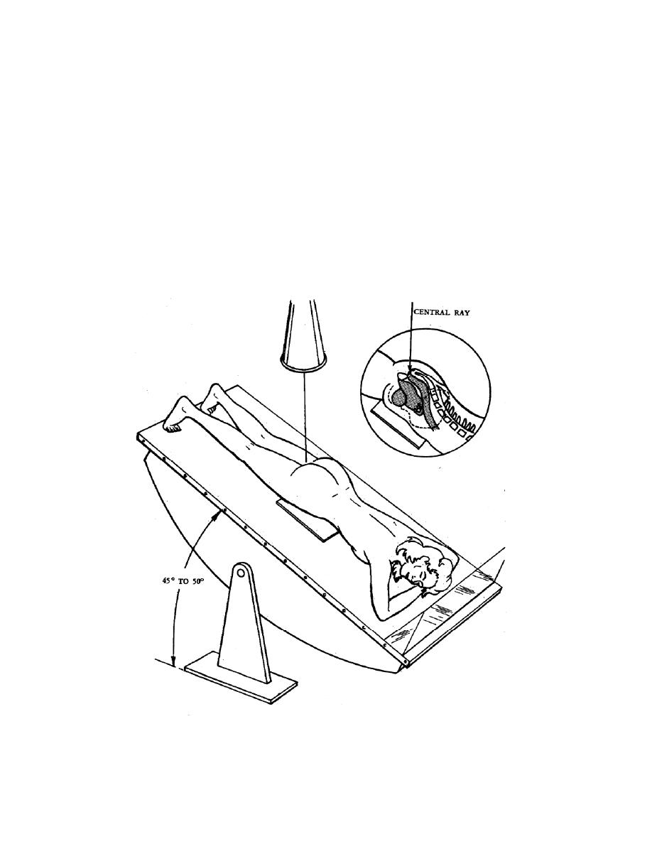
(9) Sometimes a pelvic pneumoperitoneum exposure may be performed to
demonstrate the external contours of the uterus and adjacent structures. For this
procedure, approximately 500 cc of carbon dioxide or oxygen is introduced into the
peritoneal cavity. The gynecologist introduces the radiolucent contrast medium or
radiologist while the patient is in the Trendelenburg position. For radiography, the
patient is turned in the prone position, and the table is tilted 45 to 50 to bring the hips
above the level of the head (figure 2-40). The CR is projected vertically and directed
through the region of the rectum to the center of the film. The exposure mAs should be
reduced by approximately one-half of the normal. Other radiographs are taken as
ordered.
Figure 2-40. Patient positioned for pelvic pneumoperitoneum.
MD0959
2-63

 Previous Page
