
SUGGESTED STARTING TECHNIQUE
HOLDER
*CM
KVP*
MAS**
SID
GRID
CONE
Cassette
28-34
90
220
40
8:1
Film
coverage
* Measure through the plane of the iliac crests.
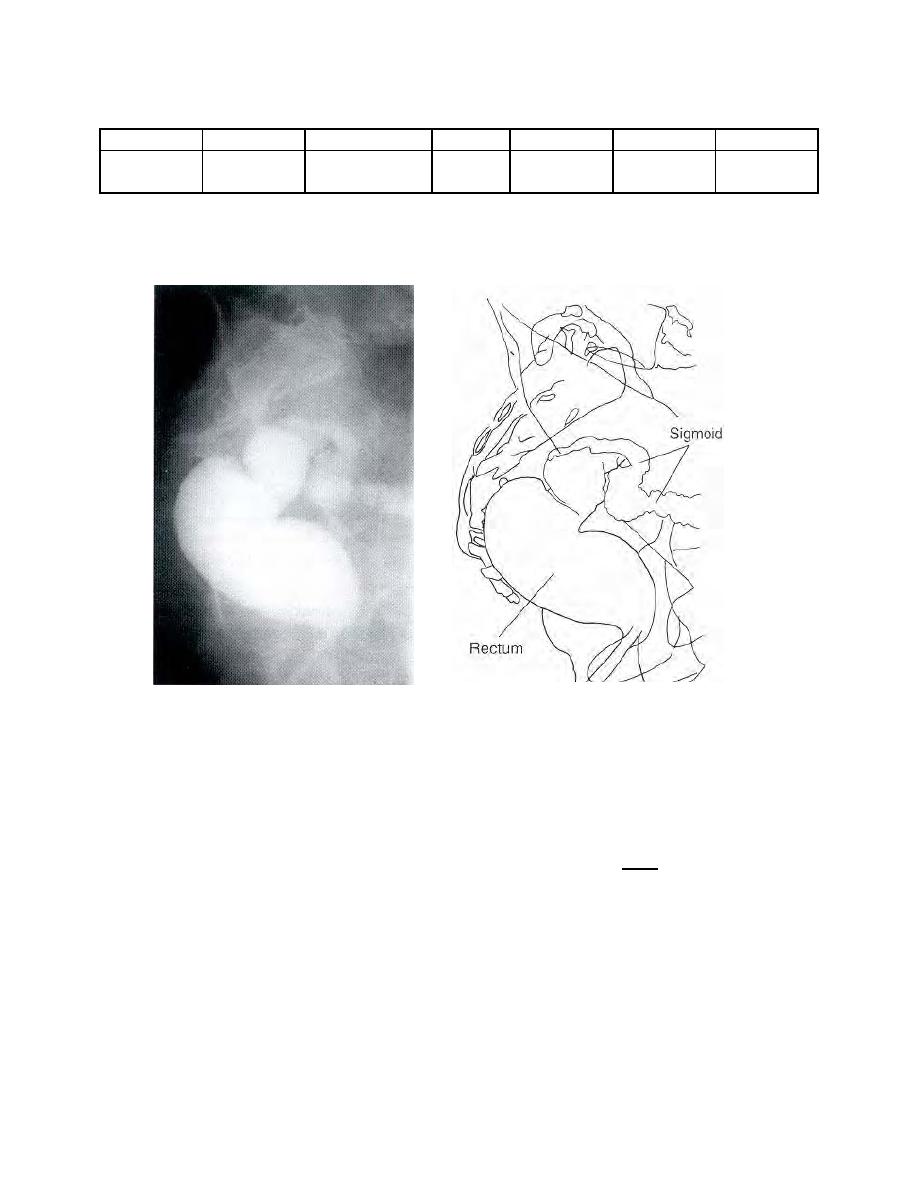
Figure 2-19. Radiograph of lateral rectum.
(b) Immediately after evacuation, the patient is recalled for fluoroscopy.
Then, air is introduced into the colon by means of a colonic insufflator. This is done
under fluoroscopic control.
(c) Routine radiographs are usually obtained in both the prone and
supine positions, because the opaque medium may tend to collect and "puddle" due to
the influence of gravity. Radiographs made with a horizontal CR and the patient in the
right lateral decubitus position and the left lateral decubitus position are often obtained.
Stereoscopic films may be made if indicated.
(d) The specialist must accomplish the necessary radiography as
rapidly as possible since the retention of a considerable volume of air in the colon may
cause distress to the patient.
MD0959
2-30

 Previous Page
