
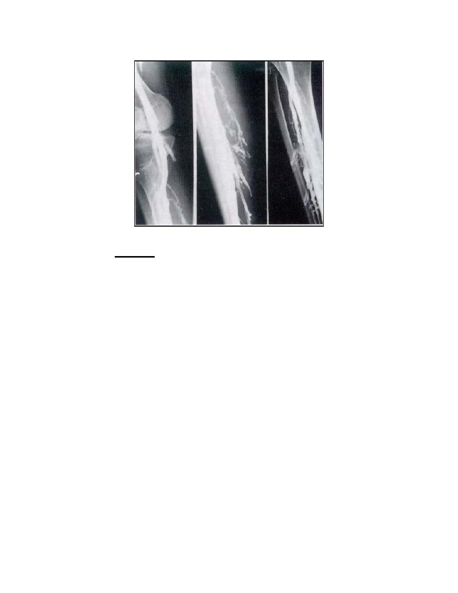
Figure 3-9. Venograms of the leg.
(4) Variations. There are numerous methods for venography of the lower
extremities. Certain variations frequently resorted to are:
(a) Stereoradiography. Since the transport of the injected contrast
material in the venous channels is relatively slow, stereoscopic projections are practical.
Stereoscopy, however, requires the use of a suitable film-changing tunnel or the Bucky
technique. The tube-shift should be made in a crosswise direction in relation to the long
axis of the part under consideration.
(b) Projections. Because of superimposition of the tibia and fibula with
certain venous channels, oblique projections (made with the leg rotated either internally
or externally as directed by the examiner) or laterals may be especially indicated.
(c) Coverage. In certain cases, the first exposure is made to
demonstrate the most distal portion of the extremity; subsequent exposures are made
with each cassette positioned progressively nearer the regions, which lie proximally.
Since the timing of these exposures and the order in which they are made is an
extremely important factor, the specialist must carry out the instructions of the examiner
with utmost exactness.
(d) Proximal injection method. The principal object of this method is to
introduce the contrast medium into the proximal portion of the venous system of the
lower extremity. The injection may be done in one of several ways. For example, the
contrast solution is injected into either the median superficial or the lateral superficial
vein of the penis in the case of the male patient or into the superficial circumflex iliac
vein in the case of the female patient. The solution may also be introduced by direct-
needle injection of the femoral vein in the region just below the crease in the groin or by
MD0959
3-24

 Previous Page
