
LESSON 2
POSITIONING FOR EXAMS OF THE PELVIC AREA
Section I. PROJECTIONS OF THE HIP
ANTERIOR POSTERIOR PELVIS
2-1.
a. Pelvic/Hip Injury. Prior to the advent of modern medicine, a fracture of the
pelvis or hip meant either spending months in recuperation or suffering a total disability.
Today, with modern techniques and materials, an orthopedic surgeon can reconstruct a
shattered hip, thus enabling the patient to lead an active life in a comparatively short
time. It all begins with a proper diagnosis, and that is done by means of X-rays. Be
sure to handle the patient carefully in any suspected pelvic or hip injury. Take your
exposures correctly the first time since the patient will probably be in great pain.
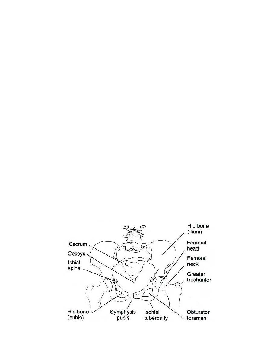
b. A Hip Routine. A hip routine consist of two views: an AP pelvis and a lateral
frog-leg hip. Additional views, such as the AP hip and the axiolateral hip (trauma), may
also be required.
c. The Anterior Posterior Pelvis. The AP pelvis is included as part of the hip routine so
that a comparison can be made between the affected side and the unaffected side. It should
be noted that the AP pelvis and the AP hip are essentially the same position. There is,
however, a difference in the film dimensions for the two positions. For the AP pelvis,
the film used is 14 x 17, so that both sides of the pelvis may be viewed on the film
simultaneously.
d. The Anterior Posterior Hip. For the AP hip, a 10 x 12 film is exposed to demonstrate
only one side of the pelvis.
MD0962
2-2

 Previous Page
