
Section IV. MOUNTING AND FILING/DISPOSING OF RADIOGRAPHS
3-21. GENERAL
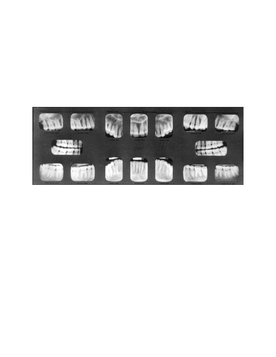
Cardboard or plastic mounts for 16-film, full-mouth radiographs and bite-wing
mounts are available as standard items of dental supply. Sections of these, or small
paper envelopes, are used for protecting and identifying individual periapical or bite-
wing radiographs. The film mounts are designed so that the film may be arranged in the
same order as the teeth in the mouth. Thus, mounting not only protects and labels the
radiographs, but also facilitates viewing and studying of the film, particularly in full-
mouth examinations. See figure 3-20.
Figure 3-20. Full-mouth radiographic mount.
3-22. MOUNTING
In mounting radiographs, care must be taken to avoid marks from damp or
perspiring fingers. Hands and fingers should be clean and dry. The film should be
handled only on the edge. Under adequate illumination, the radiographs are removed
one at a time from the hanger and placed carefully into the appropriate opening in the
film mount. Radiographs are mounted so that the raised part of the embossed dot faces
the dental specialist. In this way the radiographs are viewed from the facial aspect in
correct anatomical order.
a. Maxillary and mandibular radiographs may be identified by the anatomy of the
teeth and surrounding structures. (See paragraphs 3-24 through 3-28 for anatomic
landmarks.) Radiographs are mounted with apices of maxillary teeth directed upward
and apices of mandibular teeth directed downward.
b. The mesial aspect of a radiograph may also be determined by the anatomic
features of tissues included on the film. If the mesial is to the right (when viewed from
the facial side), it is a film taken on the patient's right side. If the mesial is to the left
(when viewed from the facial side), it is a film taken of the patient's left side.
MD0512
3-18

 Previous Page
