
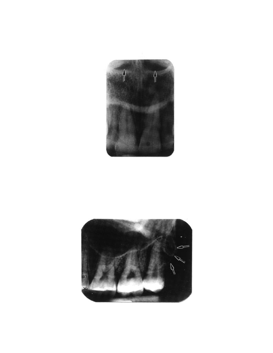
d. Nasal Fossae. In a radiograph of the maxillary central incisors, the images of
the paired fossae appear as somewhat elliptical radiolucent areas of various sizes
separated by a radiopaque band representing the nasal septum (see figure 3-24).
Figure 3-24. Nasal fossae.
3-26. RADIOPAQUE LANDMARKS ON MAXILLARY RADIOGRAPHS
a. Maxillary Tuberosity. The maxillary tuberosity (see figure 3-25) is the
convex distal inferior border of the maxilla, curving upward from the alveolar process
and distal of the third molar. An extension of the maxillary sinus is occasionally seen
within the maxillary tuberosity.
Figure 3-25. Maxillary tuberosity.
MD0512
3-21

 Previous Page
