
(7) Acanthocytes. Acanthocytes are irregularity-shaped erythrocytes with
long spiny projections. They are seen in a congenital abnormality characterized by
serum concentration of low density (beta) lipoproteins.
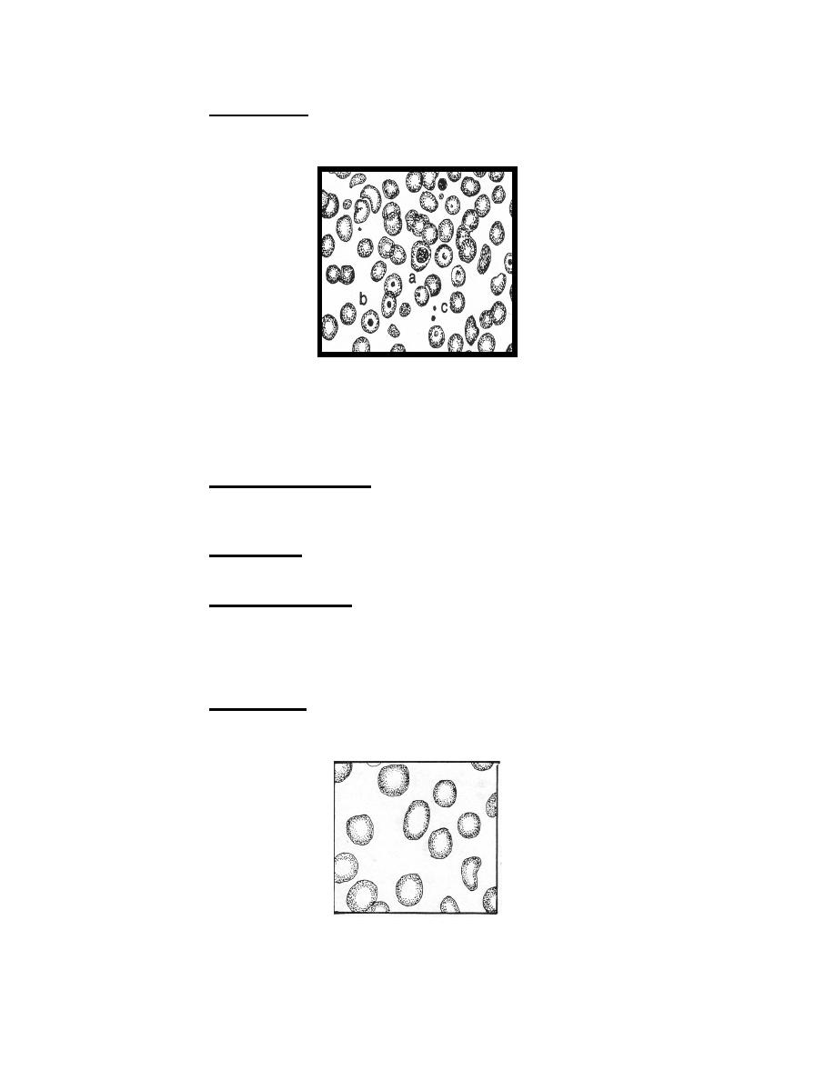
Figure 4-2f. Variations in erythrocytes:
a. Metarubricyte
b. Target Cell
c. Crenated RBC.
(8) Crenated erythrocytes. This condition occurs when blood films dry too
slowly and the surrounding plasma becomes hypertonic. There is no pathological
significance when they are found in blood smears.
(9) Schistocytes. These are red blood cell fragments. Frequently these
cells have a hemispherical shape (helmet cells).
(10) Rouleaux formation. This phenomenon is adherence of erythrocytes to
one another presenting a stack-of-coins appearance. It occurs in conditions
characterized by increased amounts of fibrinogen and globulin.
c. Staining.
(1) Hypochramia. Hypochramia is a condition in which the normal central
pallor is increased due to decreased hemoglobin content. This condition is
characteristic of many anemias.
Figure 4-2g. Variations in erythrocytes:
Hypochromic macrocytic erythrocytes.
MD0853
4-9

 Previous Page
