
e. Megaloblastic Erythrocytes. The development of megaloblastic cells is
caused by a deficiency of vitamin B12 or folic acid. Pernicious anemia is a disease
considered to be due to a deficiency in vitamin B12 and/or certain related growth factors.
With this deficiency, the erythrocytes do not mature normally and are generally larger
than normal. The most notable characteristic of this abnormal maturation is a difference
in the rates of maturation of the cytoplasm and the nucleus. The development of the
nucleus is slower than that of the cytoplasm, so that in the more mature of the nucleated
forms a spongy nucleus as well as an exceptionally large size may be observed.
Nuclear chromatin in the megaloblast is much finer and is without the clumps observed
in the rubriblast. Such development is termed asynchronism. The mature cell is large
(about 10 microns) and is termed a megalocyte. The younger cells of this series are
named by adding the suffix "pernicious anemia type," that is rubricyte, pernicious
anemia type, and so forth.
Section III. LEUKOCYTES
4-6.
GRANULOCYTIC SERIES
The stages in the normal maturation of the granulocytes are: myeloblast,
promyelocyte, myelocyte (neutrophilic, eosinophilic, and basophilic), metamyelocyte
(neutrophilic, eosinophilic, and basophilic), band cell (neutrophilic, eosinophilic, and
basophilic), and segmented cell (neutrophilic, eosinophilic, and basophilic). As the
granulocytes mature, the granules increase in number. These granules later become
specific and differ in the affinity for various dyes. Neutrophilic granules do not stain
intensely with either dye. Basophilic granules have an affinity for the basic or blue dye.
Eosinophilic stain red with an affinity for the acid dye. The criteria for identification of
the various stages of the granulocytic series are: size of cell, nucleus-cytoplasm ratio,
nuclear shape, number of nucleoli, and the type and size of cytoplasmic granulation.
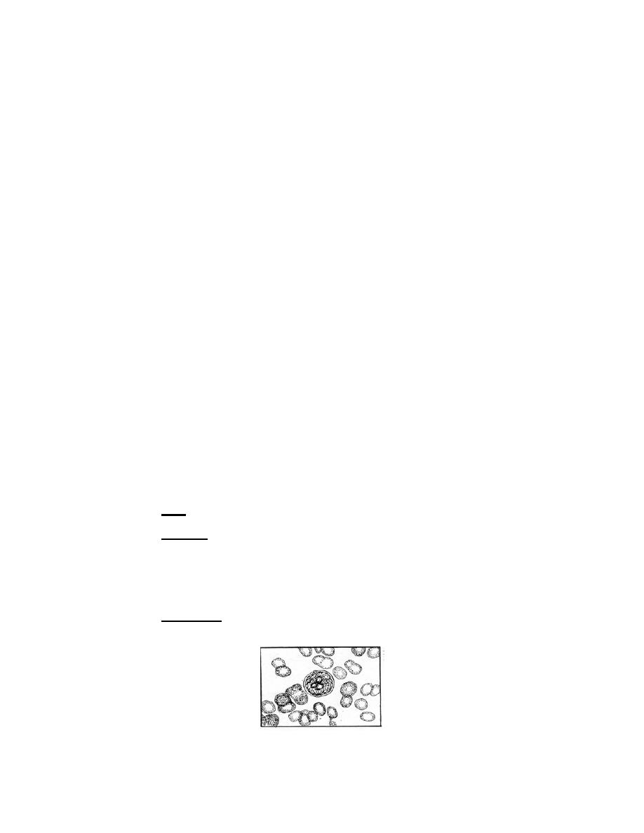
a. Myeloblast.
Size. Fifteen to 20 microns in diameter.
(1)
(2) Nucleus. The nucleus is round or ovoid and stains predominantly
reddish-purple. The interlaced chromatin strands are delicate, well defined, and evenly
stained. Two or more pale blue nucleoli are demonstrable. The nucleus occupies most
of the cell with a nucleus-cytoplasm ratio of 6:1. It is separated from the cytoplasm by a
definite nuclear membrane.
(3) Cytoplasm. The cytoplasm is a narrow, deep blue, nongranular rim
around the nucleus.
Figure 4-3a. Granulocytic series: Myeloblast.
MD0853
4-12

 Previous Page
