
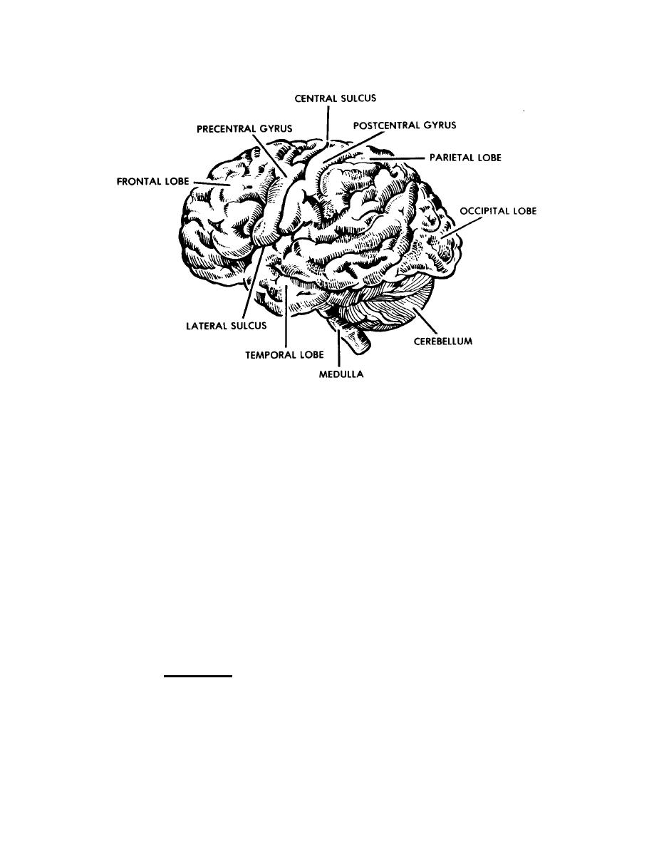
Figure 2-2. Human brain, lateral view.
(2) More centrally located within the cerebellum is the "white matter." White
matter is actually the myelin covered processes of the neurons.
(3) The cerebellum is concerned with coordination of nerve impulses to the
voluntary muscles and with the equilibrium of the body.
c. The Cerebrum. The cerebrum (figure 2-2) is the largest part of the brain.
The outer layer of the cerebrum is called the cerebral cortex. It is composed of the cell
bodies of neurons and is often referred to as the "gray matter." Many folds (gyri) and
grooves (sulci) increase the surface area of this layer. Beneath the cortex lies the
"white matter," actually the myelin covered processes of the neurons. The cerebrum is
divided into right and left hemispheres by a longitudinal fissure. A band of nerve fibers
called the corpus callosum connects the hemispheres and provides for communication
between them. Each cerebral hemisphere is further divided into lobes, which are
named after the overlying cranial bones (frontal, parietal, temporal, and occipital).
(1)
Frontal lobe.
(a) The frontal lobe is located beneath the frontal bones of the skull. It
is divided from the parietal lobes by the central sulcus and from the temporal lobes by
the lateral fissure.
MD0919
2-6

 Previous Page
