
2-4.
THE SPINAL CORD
a. The spinal cord, located within the vertebral canal of the spine, is continuous
with the brainstem. (Together, the brain and spinal cord are referred to as the
neuraxis.) The spinal cord extends from the foramen magnum of the skull to the level of
the first lumbar vertebrae, at which point it tapers to fine threads of tissue.
b. The spinal cord has two enlargements along its length that are due to an
increase in the mass of nervous tissue required to serve the limbs.
(1)
The cervical enlargement is associated with the nerves of the upper
extremities.
(2)
The lumbosacral enlargement is associated with the nerves of the lower
extremities.
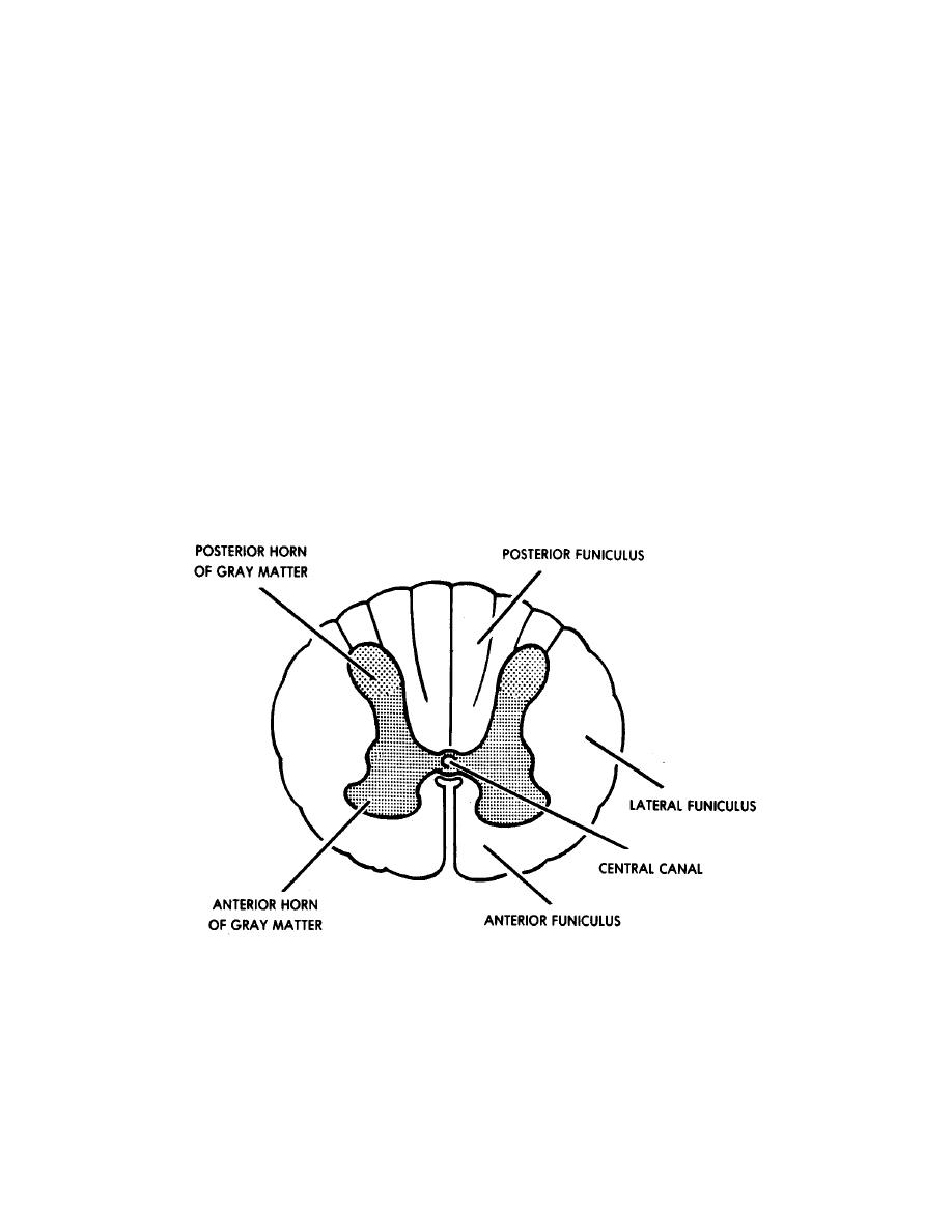
c. The spinal cord is composed of a central mass of gray matter (cell bodies of
neurons) surrounded by white matter (myelinated processes of the neurons). The areas
of gray and white matter are referred to as columns.
Figure 2-3. The spinal cord, cross-section.
d. In a cross-section of the spinal cord (figure 2-3), the gray matter appears to
be H-shaped. Each arm of the H is called a horn, resulting in two anterior horns and
two posterior horns. The connecting middle portion is called the gray commissure.
MD0919
2-8

 Previous Page
