
(5) A scalpel and heavy scissors may be used to cut through the
lumbocostal ligaments. The rib is grasped with an ochsner clamp and cut with rib
shears, removing the portion necessary to expose the kidney.
(6) Retractors and pads are placed. Gerota's fascia, the perirenal capsule,
is grasped with long tissue forceps and incised with a scalpel. The incision is extended,
using dissecting scissors, and the kidney and perirenal fat are exposed. The kidney is
dissected free, using sharp and blunt dissection with long tissue forceps, scissors, and
sponges on forceps. Crile hemostats are used on bleeding vessels.
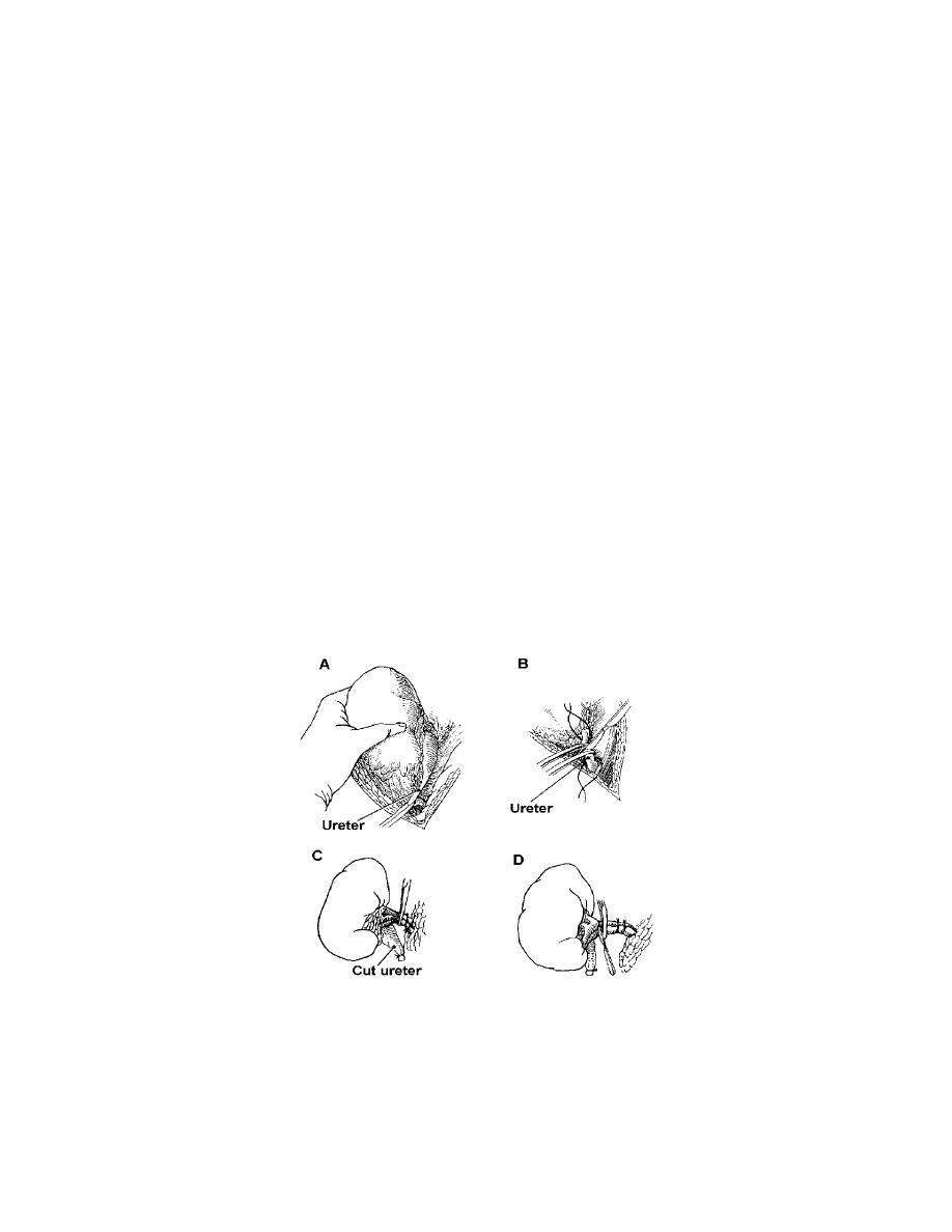
(7) The ureter is identified, separated from its adjacent structures, and
retracted. Holding forceps such as long Babcock or long Allis clamps may be used, or a
length of Penrose tubing may be passed around the ureter to retain and retract it. The
ureter is occluded by double clamping and then divided and ligated (see figures 3-6 A
and B).
(8) The kidney pedicle containing the major blood vessels is isolated and
doubly clamped by using long kidney clamps of a size suitable to the structures. The
vessels are securely ligated with heavy chromic gut and transfixed with heavy sutures
on Atraumatic needles. The pedicle is severed and the kidney removed (see figures 3-6
C and D).
(9) The wound is explored for bleeding, hemostasis secured, and the cavity
cleansed by irrigating, sponging, and suctioning as necessary. A drain of Penrose
tubing, which may be wicked with gauze, or a drain made of heavy rubber or plastic
tubing is placed if leakage of urine is likely to occur.
Figure 3-6. Nephrectomy.
A and B-Upper portion of ureter is freed, cut, and ligated. C-Chromic gut ligatures are
placed; kidney clamps are applied between proximal ligature and kidney itself.
D-Renal vascular pedicle is doubly ligated with suture ligatures, and the kidney is
removed.
MD0928
3-12

 Previous Page
