
to in radiography as the petrous ridge. The level of the petrous ridge (figure 2-31) is
about 1 inches above the apex of the mastoid tip. On the posterior surface near the
center is a large orifice, the internal acoustic meatus. Within the petrous portion are the
auditory ossicles (the malleus, the incus, and the stapes), the cochlea, and the
semicircular canals. The auditory ossicles and the cochlea are essential portions of the
hearing mechanism and the semicircular canals are concerned with equilibrium.
(5) The styloid process (figure 2-30) is a slender, pointed, process attached
to the inferior surface of the tympanic portion.
d. Occipital. The occipital bone (figures 2-28, 2-29, and 2-30) is at the posterior
and inferior portion of the cranium. A large hole, the foramen magnum (figures 2-30
and 2-31), pierces the occipital bone. The occipital bone is divided into four parts: the
squama (behind the foramen magnum), the basilar portion (anterior to the foramen),
and two lateral portions, one on each side of the foramen magnum. The curved
expanded squama presents externally the external occipital protuberance (EOP).
Situated on the internal surface of the squama, which is deeply concave, are the
internal occipital protuberance and the grooves which lodge the blood sinuses. On the
inferior surfaces of the lateral portions are the occipital condyles that articulate with the
atlas (C-1) of the spine. The basilar portion extends anteriorly and superiorly from the
foramen magnum and is attached to the body of the sphenoid bone.
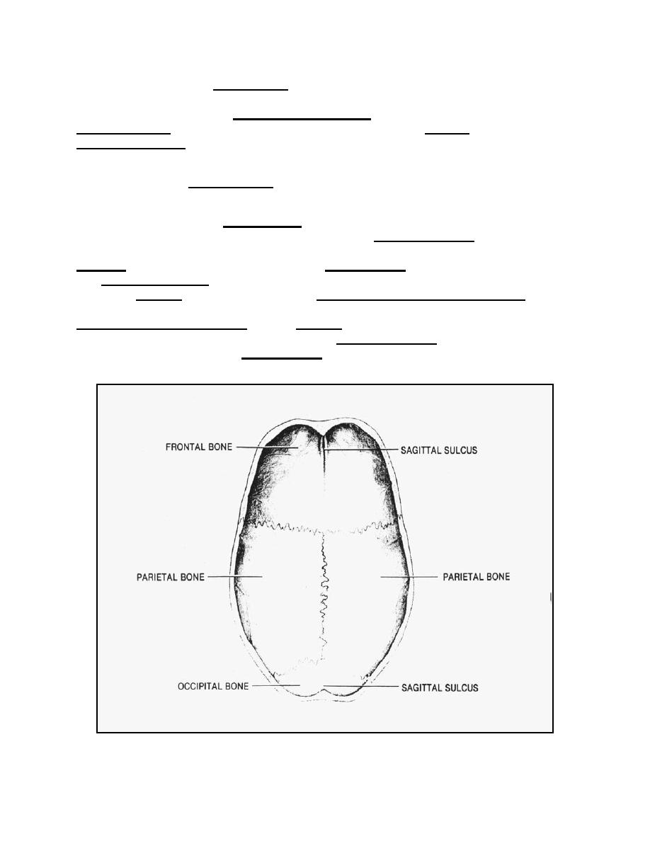
Figure 2-32. Inner surface of the cranial roof.
MD0956
2-48

 Previous Page
