
e. Sphenoid. The sphenoid bone (figures 2-27, 2-28, 2-29, and 2-30) is at the
base of the skull in front of the temporals and the basilar part of the occipital bone. In
form, it resembles a bat with its wings extended. It is divided into a body, two greater
wings, two lesser wings, and two pterygoid processes.
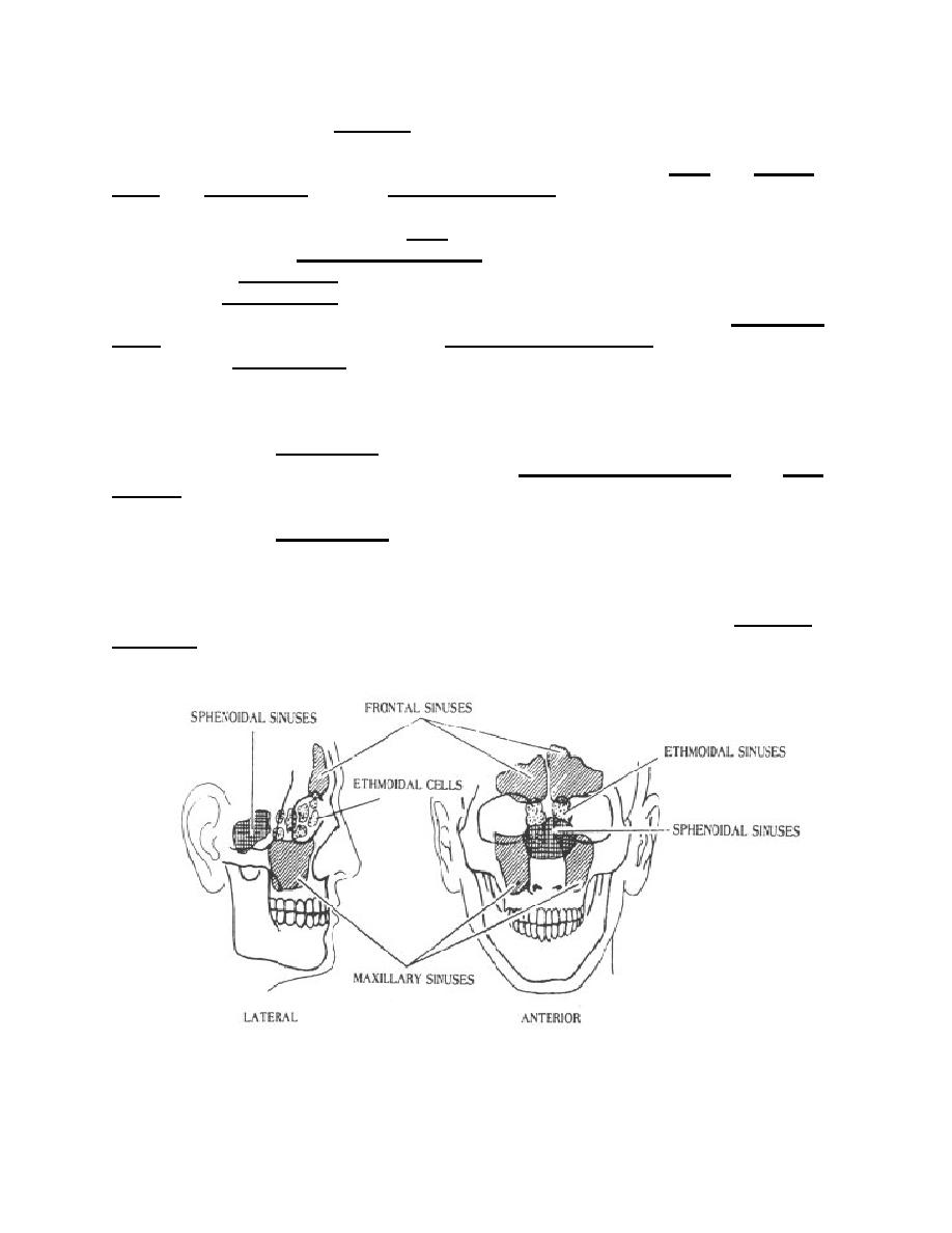
(1) The cubical-shaped body (figure 2-31) is hollowed out internally to form
two large cavities, the sphenoidal air sinuses (figure 2-35). On the superior surface of
the body is the sella turcica (Turkish saddle) (figures 2-28 and 2-31). Situated within the
saddle is the pituitary fossa, that receives the pituitary gland. Anteriorly, the sella
turcica is bounded by an eminence, the tuberculum sellae. Anterior, to the tuberculum
sellae, is a transverse groove called the optic(chiasmatic) groove, which terminates
laterally in the optic foramen. Each optic nerve leaves the posterior aspect of the eye,
goes through the optic foramen, crosses in the chiasmatic groove (one-half of the fibers
do not cross), and terminates in the brain.
(2) The lesser wings (figure 2-31) project laterally away from the anterior
aspect of the body and terminate medially as the anterior clinoid processes. The optic
foramen, situated in the back of the orbit, is part of the lesser wing.
(3) The greater wings (figure 2-31) project anteriorly and laterally away from
the body to form, in part, the posterior aspect of the orbit and a portion of the lateral
walls and floor of the cranium.
(4) The "legs" of the bat descend from the greater wings as the pterygoid
processes (figure 2-28).
Figure 2-35. The paranasal sinuses (lateral and anterior aspects).
MD0956
2-51

 Previous Page
