
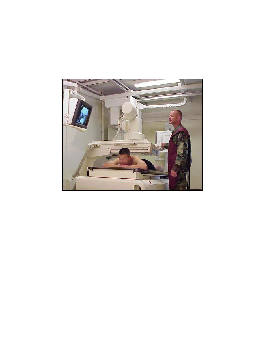
(a) Patient prone (figure 3-17). The patient is adjusted in the prone
position with the midline of the body aligned to the center of the table; the head may be
turned to either side. The patient is then instructed to grasp the edge of the tabletop
with his hands at the level of his shoulders or to grasp the shoulder brace with one
hand. To straighten the lumbar curve to the required degree, a bolster (made by rolling
a small pillow or other suitable object into the desired shape) is placed under the lower
abdomen. The patient's feet are placed firmly against the footrest.
Figure 3-17. Head-end of tilt-table unit raised during myelographic examination.
(b) Patient lying on his side (figure 3-18). The patient lies on his side,
with his back toward the examiner. A suitable bolster is placed under the thoracic
region to straighten the spinal column in relation to the midsagittal plane and make it
parallel with the tabletop. The patient's neck, body, and lower extremities are then
brought into flexion so that the knees are drawn toward the chin and arms and
shoulders are drawn forward.
(3)
A sterile-packed myelographic layout is set up near the examiner.
(4)
The injection site is made aseptic and local anesthesia is administered.
(5) The radiographic and fluoroscopic factors are determined according to
the size of the patient. If necessary, test exposures are made and developed for
immediate inspection. The specialist should prepare the radiographic room for the
examination so that every item needed will be in place. A sufficient number of films of
the required sizes should be available both for spot-filming and for other radiography.
Appropriate identification markers should be made before any radiography is done.
MD0959
3-48

 Previous Page
