
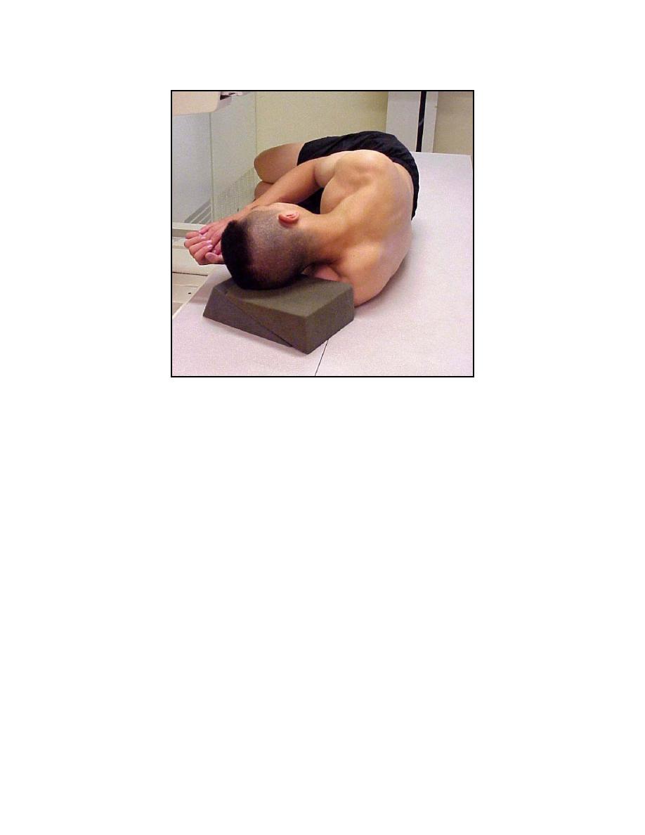
Figure 3-18. Patient lying on his side, body in flexion, for myelography.
d. Introduction of Contrast Medium, Fluoroscopy, and Radiography.
(1)
Sterile and aseptic precautions are observed throughout the entire
procedure.
(2) The examiner inserts an 18- or 20-gauge lumbar-puncture (short-
beveled) needle into the subarachnoid space exactly in the midline and in the region of
the vertebral interspaces between L3 and L4, L4 and L5, or L5 and S1. The stylet is
removed from the needle and approximately 3 to 5 cc of cerebrospinal fluid are
withdrawn and collected in a test tube for laboratory analysis. When indicated by the
examiner, the Queckenstedt test is performed for the determination of possible block in
the vertebral canal. In the absence of an assistant, the examiner may direct the
specialist to assist him in this maneuver (carotid artery compression). In this case, the
specialist must carry out the examiner's instructions (however simple they may seem)
with exacting care. The cerebrospinal fluid pressure is recorded at this stage of the
procedure.
MD0959
3-49

 Previous Page
