
2-4.
SMALL INTESTINE (SMALL BOWEL SERIES)
In the investigation of the small intestine, the study is usually done by a
combination of fluoroscopic and radiographic methods. The preparation of the patient,
contrast medium, and management of the facilities are essentially the same as for the
examination of the stomach. The radiologist may direct that ice-cold normal saline
solution (cold isotonic method) be used as the vehicle for the barium sulfate in place of
water, to speed up the examination. The cold solution stimulates peristalsis, causing
the barium to pass more rapidly through the gastrointestinal tract. In the double
method, a designated quantity of contrast medium is administered to the patient at a
specified time prior to fluoroscopy; during fluoroscopy, additional barium is given to the
patient (spot-filming may also be done at the time). Radiographs are made at the
discretion of the radiologist: for example, a film every 15 minutes for the first hour, then
at half-hour intervals, as indicated. Appropriate identification markers should be used
for each exposure to indicate the time intervals.
2-5.
LARGE INTESTINE (DOUBLE CONTRAST) BARIUM ENEMA
a. A method that is widely used for the introduction of the contrast media into the
colon is based on a double contrast consisting of barium and air. The liquid component
of the contrast media is introduced into the colon by means of gravity. Once the barium
has coated the lining of the colon, the barium is mostly drained out of the colon before
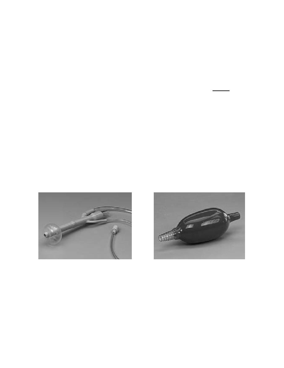
air is administered. Using the Air-Contrast enema tip and inflator bulb (figure 2-12), air
is slowly pumped into the colon either by the radiologist or per his instructions.
Figure 2-12. The rectal tip with catheters for inflation of a retention balloon and
an inflator bulb used to inflate the balloon and administer air for
double-contrast examinations.
MD0959
2-17

 Previous Page
