
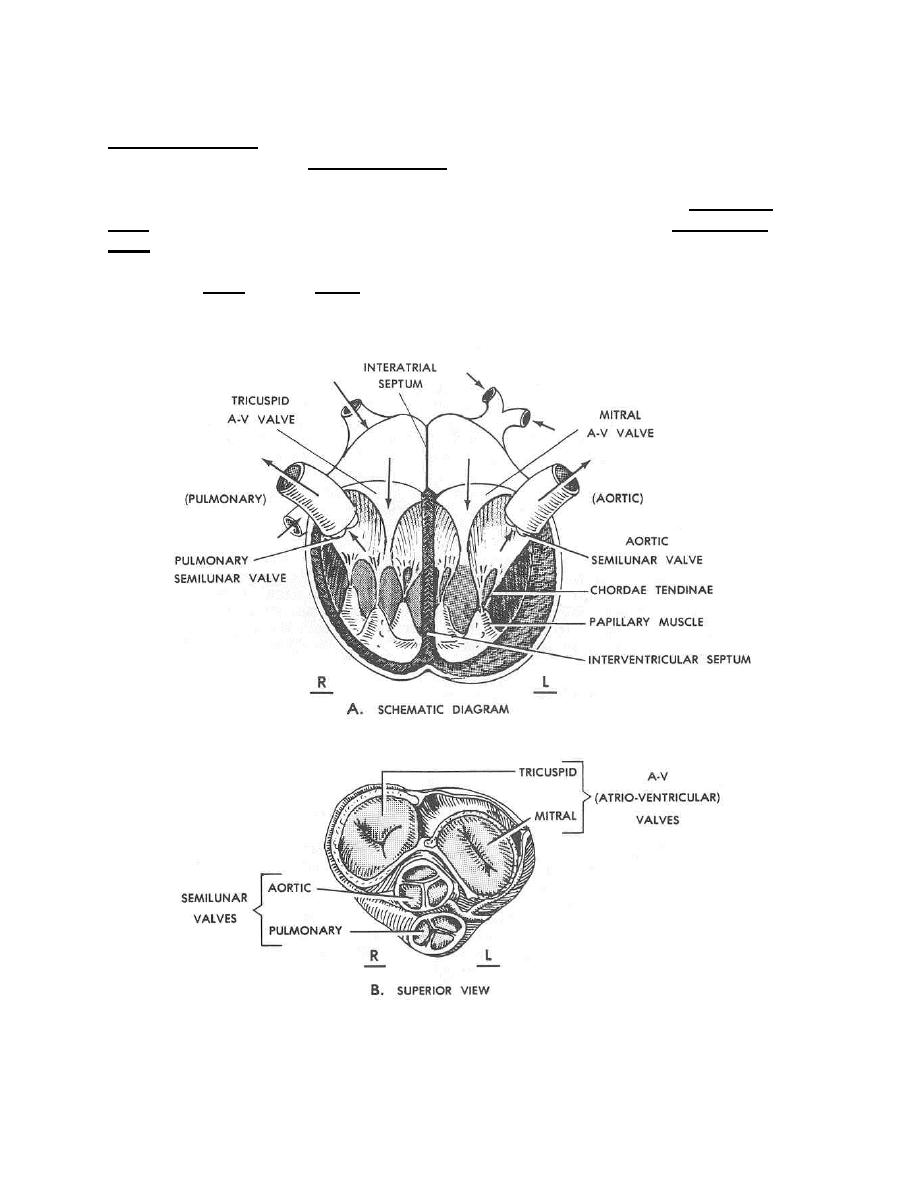
cords are attached to the undersides (the ventricular side) of the leaflets and are called
chordae tendineae. At their other ends, the chordae tendineae are attached to the inner
walls of the ventricles by papillary muscles.
(b) A major artery leads away from each ventricle: the pulmonary
trunk from the right ventricle and the aortic arch from the left ventricle. A semilunar
valve is found at the base of each of the pulmonary trunk and the aortic arch. These
semilunar valves prevent blood from flowing back into the ventricles. The pulmonary
(semilunar) valve and the aortic (semilunar) valve are each made up of three semilunar
("pocket-like") cusps.
Figure 2-3. Scheme of heart valves.
MD0806
2-16

 Previous Page
