
3-26. RADIATION THERAPY TREATMENT PLANNING
The computer can be used to produce a precise radiation therapy treatment plan.
The technologist simply keys in the relevant patient information, such as, diagnosis,
condition, and overall treatment. The computer will then generate a printout specifying
type, number, and schedule of treatments.
3-27. NUCLEAR MEDICINE APPLICATIONS
This technology uses radioactive isotopes and computerized scanners to provide
accurate determinations about the presence or absence and nature of abnormalities. In
liver or brain scans, for example, a radioisotope is injected into the body. The
computerized scanner detects the degree of concentration of the radioisotope energy
received from the patient. It stores this data for recall so at the radiologist can examine
the area of interest, plane by plane. Higher or lower concentrations of the radioisotope
energy in a certain area indicate the presence or absence of an abnormality, such as a
tumor.
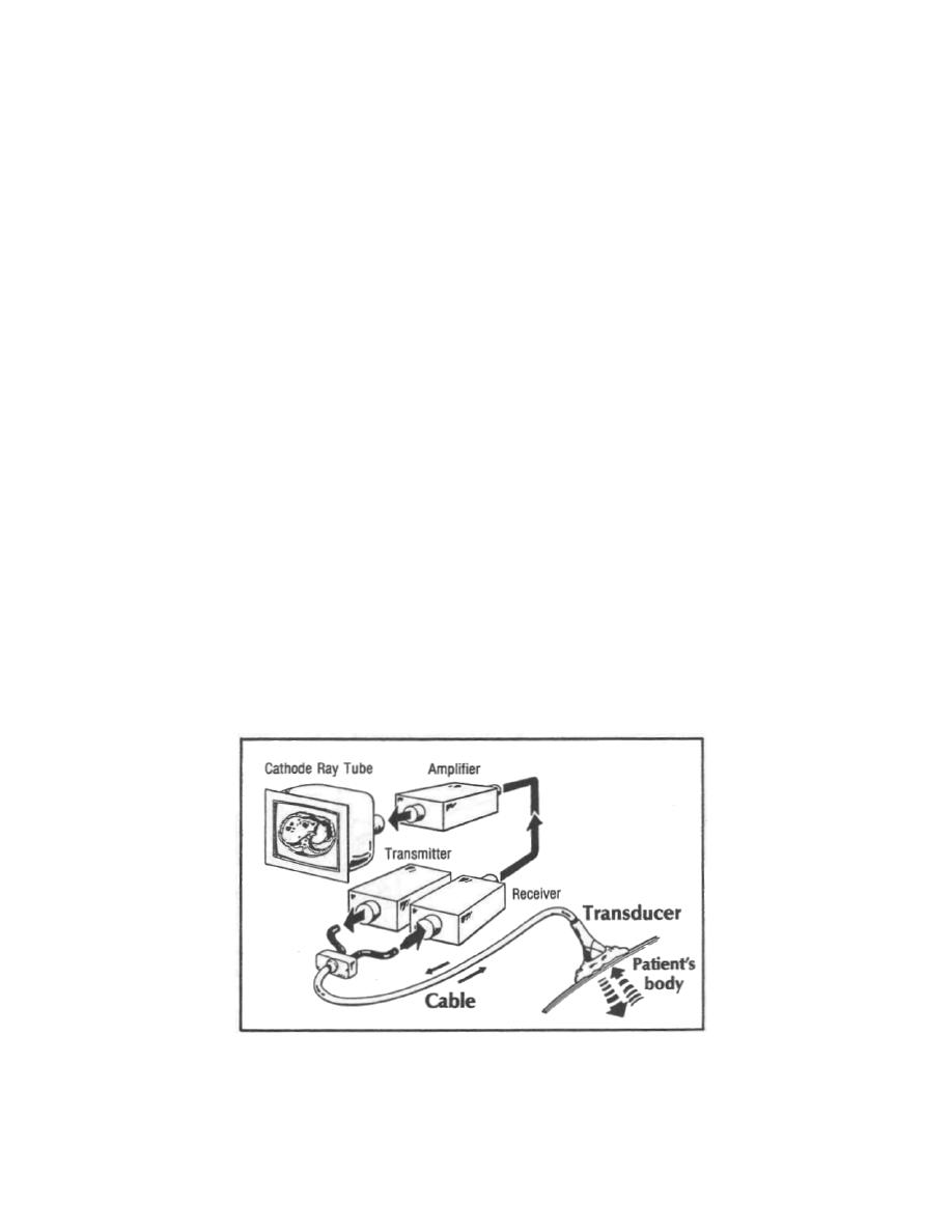
3-28. ULTRASOUND APPLICATIONS
Ultrasound technology uses the sound waves reflected by anatomical structures
to determine size and location of tumors, fetuses, and so forth. The relationship of one
echo relative to another is what helps localize the body anatomy. A device called a
transducer is placed in direct contact with the patient. With its flexible cord arms, the
transducer can accurately follow the contours of the body without repositioning the
patient. The transducer produces and receives sound waves that a small computer
stores and recalls. The sound waves are used in calculating the alignment of the
echoes relative to each other, which determines size and location of the anatomic
structures.
Figure 3-12. Using the transducer, a view of the abdomen is produced on
the CRT screen. The dark round spots on the liver (large gray
mass on the left) may signal a tumor.
MD0058
3-21

 Previous Page
