
1-50. LEUKOPLAKIA
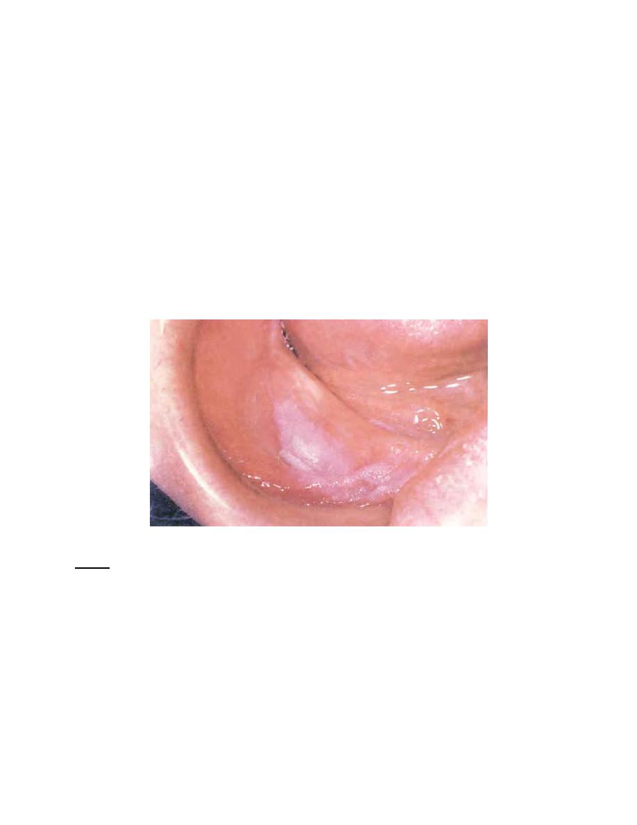
Leukoplakia (figure 1-13) is a clinically descriptive term defining a white plaque.
The white plaque is observed as irregular, thickening of the outer layers of the mucosa,
a dull-white-to-gray color, variable size, and with very little pain unless ulceration and
secondary infection have developed. The predominate oral cause is commonly
associated with chronic irritation or trauma, such as wearing of ill-fitting dentures,
cheekbiting, oral use of tobacco, or malpositioned or rough tooth surfaces. These areas
are generally discovered by the dentist on routine examination, the patient being
unaware of their existence. The condition may precede development of a malignant
tumor. For this reason, early diagnosis and treatment is important. Leukoplakia
resembles other conditions that may affect the oral mucosa; therefore, diagnosis must
be based upon microscopic examination of cellular changes within the involved tissues.
Tissue specimens are prepared for examination by a procedure called biopsy. Once a
definitive diagnosis has been made, the treatment of choice may be removal of the
lesion.
NOTE: This white plaque has formed in an area commonly associated with denture
wear. Any area of leukoplakia that does not return to normal within 10 to 14
days after removal of the cause of irritation must be biopsied to confirm a
benign cellular change.
Figure 1-13. Leukoplakia.
MD0511
1-26

 Previous Page
