
b. Coronoid Process of the Mandible. The coronoid process of the mandible
(see figure 3-26) sometimes appears on maxillary molar films as a triangular opaque
area located in the region of or distal to the maxillary tuberosity.
Figure 3-26. Coronoid process of the mandible.
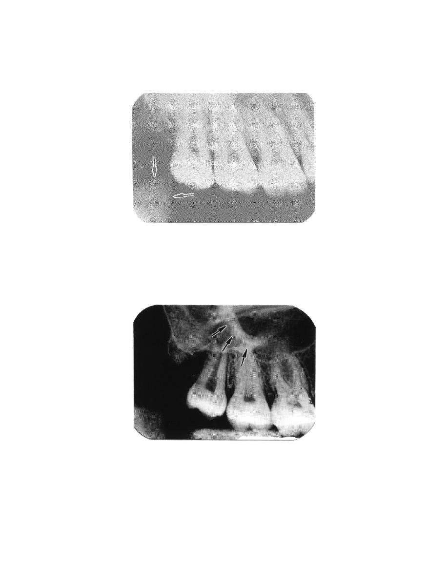
c. Zygomatic Process (Malar Bone). The zygomatic arch (see figure 3-27)
commonly appears as a well-defined radiopaque area that may be superimposed over
the molar roots. Additional radiographs are sometimes made at adjusted angulation to
provide a better view of the molar root area.
Figure 3-27. Zygomatic process (malar bone).
d. Nasal Septum. The nasal septum is usually seen as a white ridge extending
above and between the central incisors.
MD0512
3-22


 Previous Page
