
4-13. MAXILLARY INCISORS
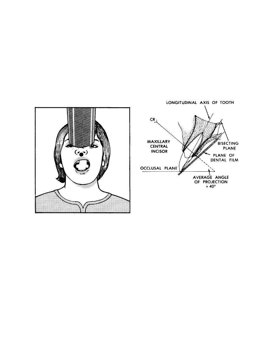
Adjust the head as described for radiography of maxillary teeth. Gently contour
both long borders of the film slightly in the direction of the curvature of the palate. Place
the film packet in the mouth so that its long borders are vertical and its center is in line
with the median plane of the upper arch. Have the lower border of the packet slightly
below (approximately 1/8 inch) and parallel to the incisal edges. Adjust the tube to an
average angulation of +40 degrees. Direct the central ray to pass through the tip of the
nose in line with the median plane and perpendicular to the bisecting plane (see figure
4-13). Follow the manufacturer's instruction for exposure times.
Figure 4-13. Maxillary incisor area.
4-14. MANDIBULAR MOLARS
Adjust the head as described for radiographs of mandibular teeth (paragraph
4-7b). Place the packet in the mouth with the long axis horizontal and the upper border
of the film parallel to and slightly above (approximately 1/4 inch) the occlusal surfaces of
the molar teeth. Relieve the lower anterior border by contouring. Place the packet
alongside the tongue and far enough distally to include the entire third molar area.
Impacted or malposed mandibular teeth may require special positioning of the film
packet. Adjust the tube to an average angulation of -5 degrees. Direct the central ray
straight through the interproximal spaces at the center of the film and perpendicular to
the bisecting plane (see figure 4-14). Follow the manufacturer's instructions for
exposure times.
MD0512
4-11

 Previous Page
