
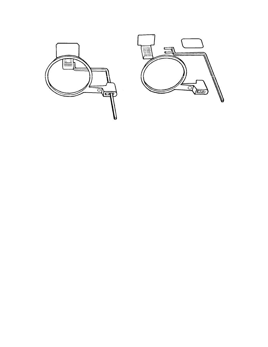
Figure 4-20. Posterior instrument assemblage.
(1) The shielded, printed, or broken side of the film packet is placed against
the backing support of the bite-block.
(2) It is inserted horizontally into the slot by using a downward motion and,
at the same time, placing slight pressure against the backing support to open the slot.
(3) The embossed dot on the corner of the film is also placed in a downward
position when placed in the plastic bite-block.
(4) The right angle portion of the indicator rod is held anterior to the bite-
block and away from the film.
(5) The pins are inserted into the proper holes. (The three holes allow a
choice for the desired lingual positioning of the film).
(6)
The plastic locator ring is fitted onto the indicator rod opposite the film
packet.
(7)
The assembly is then positioned in the mouth.
4-20. MAXILLARY MOLARS
Position the posterior instrument assembly in the patient's mouth with the plastic
bite-block centered on the second molar (see figure 4-21). Be sure that the anterior
edge of the film is adjacent to the distal of the second bicuspid. Parallel the film with the
long axis of the molars. Place a cotton roll between the underside of the teeth and the
block and have the patient close his teeth in order to maintain the film position. Move
the locator ring along the indicator rod to approximately the skin surface and align the
x-ray unit extension tube with the rod and the ring on horizontal and vertical planes.
MD0512
4-16


 Previous Page
