
appears cloudy or discolored, the probable cause is a cataract. Health of the iris is
determined by noting the regularity of the pupil. An irregular, constricted appearance to
the pupil may result from edema due to inflammation of the iris. Screen visual acuity with
any available print. If the patient cannot read the largest print, test the patient's ability to
count your upraised fingers and distinguish light (such as your flashlight) from dark.
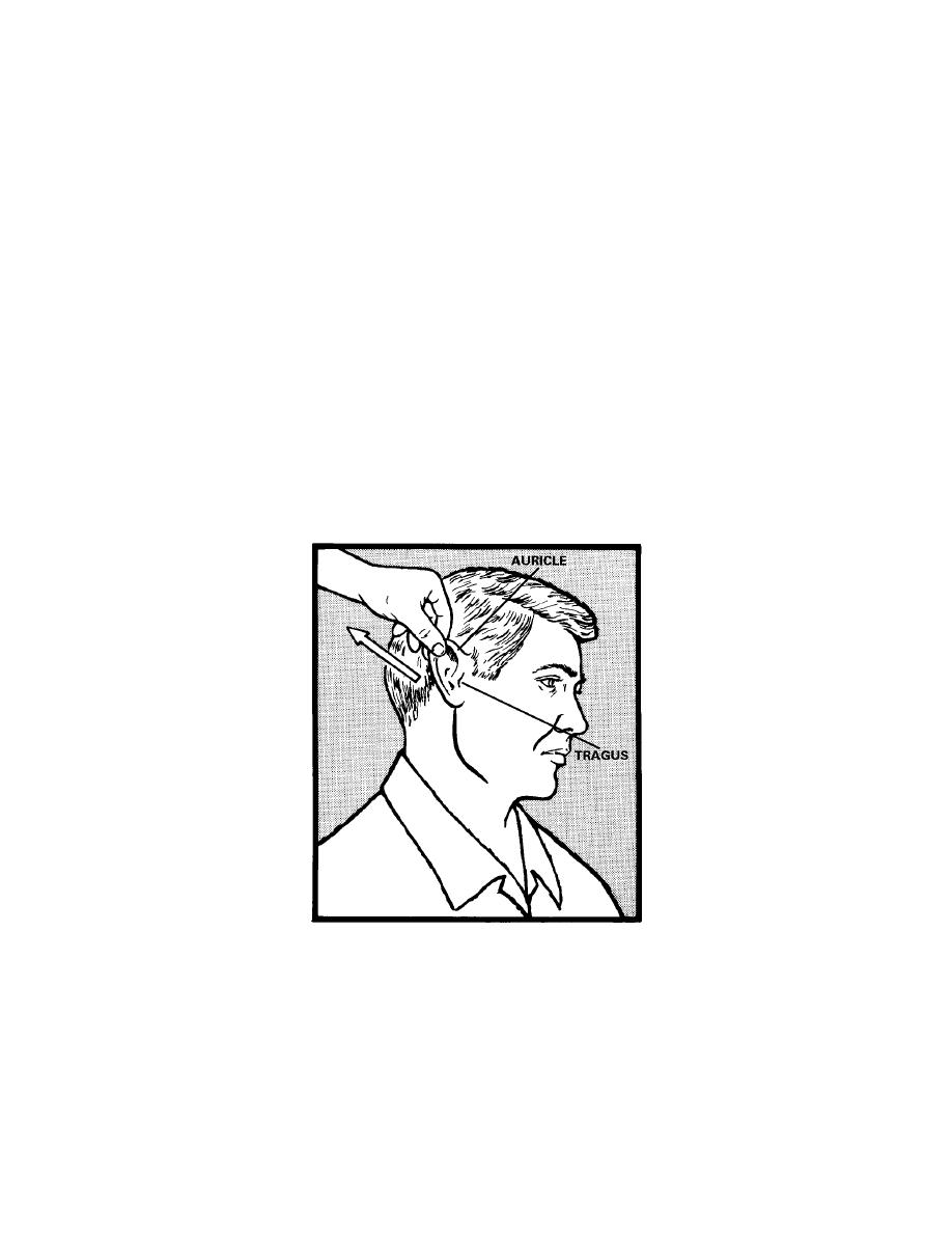
(3) The ear has three compartments: the external ear, the middle ear and the
inner ear. Much of the middle ear and all of the inner ear are inaccessible to direct
examination. The external ear is comprised of the auricle and ear canal. The ear canal
opens behind the tragus. Assess the ears for hearing, symmetry, discharge, tinnitus
(ringing in the ears), and vertigo (dizziness). Inspect each auricle of the ear and
surrounding tissue for deformities, lumps, or skin lesions. If ear pain, discharge, or
inflammation is present, move the auricle up and down, press the tragus, and press firmly
just behind the ear. Movement of the auricle and tragus (figure 6-5) is painful in acute
external otitis, but not in otitis media. Tenderness behind the ear may be present in otitis
media. To estimate hearing, test one ear at a time. Ask the patient to occlude one ear
with a finger. Stand 1 or 2 feet away, and whisper softly to the enucleated ear. Speak
words with equally accented syllables, such as "homerun" or "four-nine." Make sure that
the patient does not read your lips. Ask him to repeat what you have said.
Figure 6-5. Movement of the auricle.
(4) The nose has two major functions. It enables us to use our sense of smell
and it is the air conditioner of the respiratory system. Assess the nose for bone alignment
and epistaxis (nosebleeds). Inspect the nasal mucosa and septum. If the patient
complains of nosebleeds, ask him about the frequency, amount, and color of the
nosebleeds. Inspect and palpate the outside of the nose. By using a penlight or
MD0906
6-9
 Previous Page
