
b. Each kidney is surrounded by a mass of fatty and loose areolar tissue, known
as perirenal fat. Each kidney and fat capsule is surrounded by a sheath of fibrous
tissue called Gerota's capsule, or renal fascia, which is connected to the fibrous tunic of
the kidney by trabeculae. The kidneys are held in place by the renal fascia, which
connects with the fascia of the quadratus lumborum muscle of the loins, the psoas
major muscles, and the diaphragm.
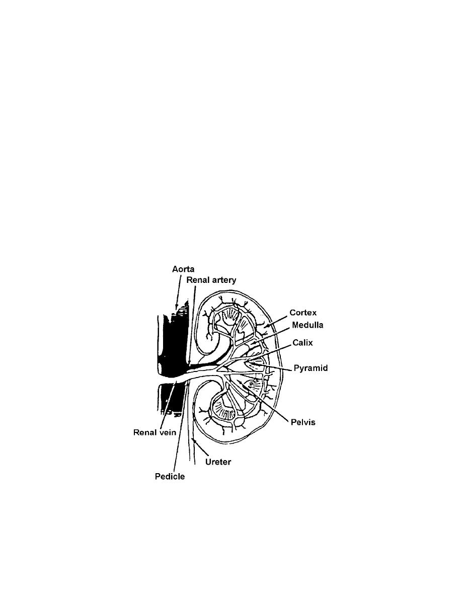
c. On the medial side of each kidney there is a concave notch (called the hilum)
through which the ureter, arteries, and veins enter and leave and where the renal pelvis
is found.
d. The substance of the kidney (see figures 3-2 and 3-3) consists of an outer
portion called the cortex, and an inner portion, called the medulla. The cortex contains
the glomeruli (see figures 3-3 and 3-4) and the functioning tubules. The medulla
contains many collecting tubules and papillary ducts. Each of the latter empties on a
papilla within a minor calyx. Several of these join to form a major calyx. These unite to
form--and therefore in turn empty into--the renal pelvis, consisting of smooth muscles
lined with epithelium. The funnel-shaped renal pelvis of each kidney is continuous with
the ureter below.
Figure 3-2. The kidney.
MD0928
3-3

 Previous Page
