
NOTE:
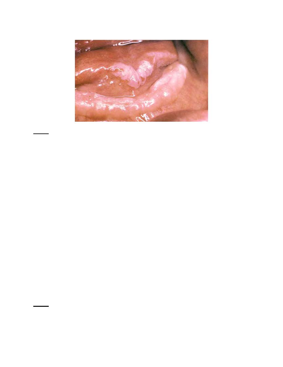
The clinical appearance of leukoplakia revealed a squamous cell carcinoma
on histologic examination. The mouth floor is a common site for oral cancer.
All oral tissues must be evaluated for abnormality with each oral examination
so that malignant growths may be recognized early in development.
Figure 2-8. Oral cancer, floor of the mouth.
2-7.
BIOPSY
a. General. Biopsy is the procedure for obtaining a tissue specimen for
microscopic examination in order to establish a diagnosis. This procedure is used when
tissue growth cannot be diagnosed by clinical observation alone. Biopsy is extremely
important in diagnosing malignant growths (tumors) requiring early treatment and in
identifying other conditions requiring early treatment or specific types of treatment.
Surgical excision (complete removal of involved tissues) or removal of a part of the
tissue may be done. Excision is the method of choice when the lesion is small and
complete removal is the treatment.
b. Processing the Specimen. After the tissue specimen has been removed
surgically, it should be placed in a container with 10 percent formalin. The container
should be large enough to accommodate fixative solution equal to about 20 times the
volume of the specimen. Usually 48 to 72 hours is enough time to ensure adequate
fixation. However, the time will be determined by the size of the specimen. The
specimen, a properly completed Standard Form 515 (Medical Record-Tissue
Examination) with five copies, a clinical history, and appropriate dental radiograph
should all be forwarded to the oral-maxillofacial pathologist.
NOTE:
The container must be properly identified in case it is separated from the
SF 515, to include the patient's name and social security number, type of
tissue sample (for example, soft tissue, bone), doctor's name, clinic address,
and date.
MD0511
2-8

 Previous Page
