
features that are not seen in other cells. Another feature of the nucleus, which is of
value in diagnosis, is the tendency for the nuclear chromatin to be loose with light
spaces in between the chromatin strands, giving a coarse linear pattern in contrast to
the Iymphocyte that has clumped chromatin. Nucleoli are absent. The nucleus-
cytoplasm ratio is approximately 3:1.
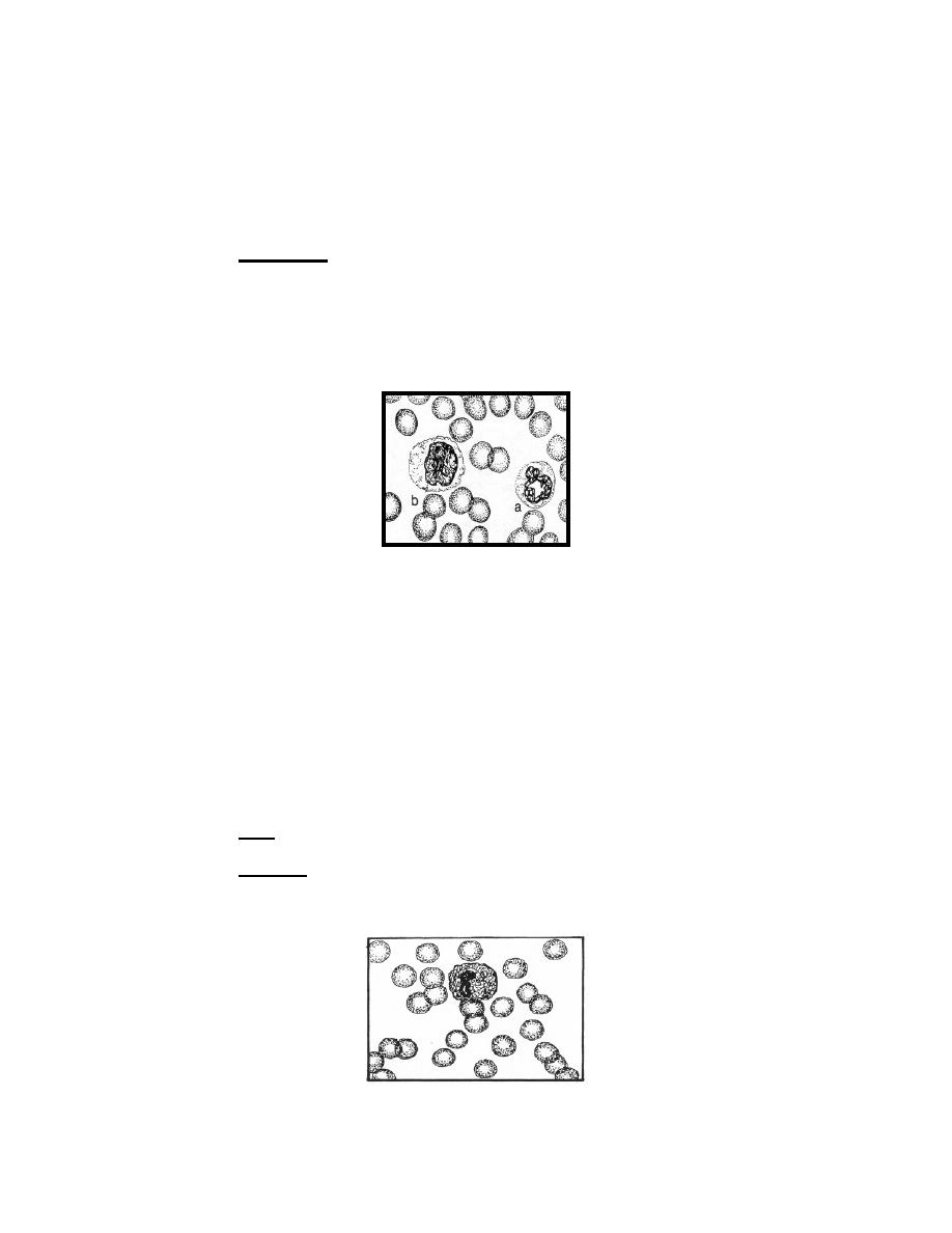
(3) Cytoplasm. The cytoplasm of the monocyte is dull gray-blue while the
cytoplasm of the neutrophils in the adjacent fields is definitely lighter and is pink rather
than gray-blue. The nonspecific granules of the monocyte are usually fine and evenly
distributed, giving to the cell a dull, opaque or ground-glass appearance. In addition to
the background of evenly distributed nonspecific granules, there may be a few unevenly
distributed larger azurophilic granules. Vacuoles are often demonstrable in the
cytoplasm.
Figure 4-5c. Monocytic series:
a. Neutrophil (Late Band)
b. Monocyte.
4-10. PLASMOCYTIC SERIES
Plasmocytes constitute approximately one percent of the white cells in the
normal bone marrow. These cells can represent in the peripheral blood in chronic
infections, granulomatous and allergic diseases, and multiple myeloma. The stages of
development are: plasmoblast, proplasmocyte, and plasmocyte.
a. Plasmoblast.
Size. Eighteen to 25 microns in diameter.
(1)
(2) Nucleus. The nucleus is large, oval or round, and located off center in
the cell. Nuclear chromatin is fine, purplish, and reticulated. There are multiple nuclei
that may or may not be visible.
Figure 4-6a. Plasmocytic series: Plasmoblast.
MD0853
4-20

 Previous Page
